Western blot analysis of extracts of HeLa and OVCAR3 cells using TIF1β (C42G12) Rabbit mAb.

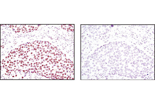
Immunohistochemical analysis of paraffin-embedded human breast carcinoma using TIF1β (C42G12) Rabbit mAb in the presence of control peptide (left) or antigen-specific peptide (right).

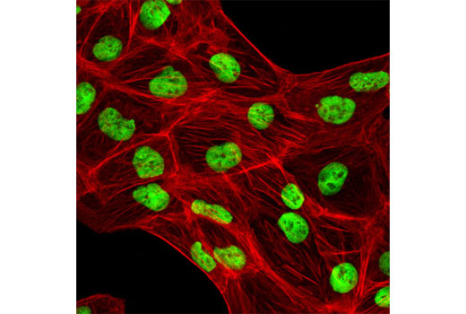
Confocal immunofluorescent analysis of ACHN cells using TIF1beta (C42G12) Rabbit mAb (green). Actin filaments have been labeled with DY-554 phalloidin (red).

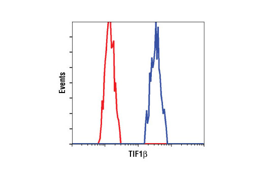
Flow cytometric analysis of HeLa cells using TIF1β (C42G12) Rabbit mAb (blue) compared to a nonspecific negative control antibody (red).


