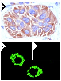
Cox-2 (M-19): sc-1747. Immunoperoxidase staining of formalin-fixed, paraffin-embedded human lung tumor showing membrane and cytoplasmic staining (A). Immunofluorescence staining of methanol-fixed RAW 264.7 cells induced with LPS and PMA showing cytoplasmic vesicle localization (B) and untreated control RAW 264.7 cells (C).

Western blot analysis of Cox-2 expression in uninduced (A,C) and LPS + PMA-treated (B,D) RAW 264.7 whole cell lysates. Antibodies tested include Cox-2 (C-20): sc-1745 (A,B) and Cox-2 (M-19): sc-1747 (C,D).
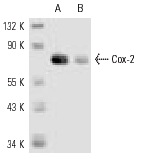
Cox-2 (M-19)-R: sc-1747-R. Western blot analysis of Cox-2 expression in NIH/3T3 (A) and LPS/PMA-treated RAW 264.7 (B) whole cell lysates.
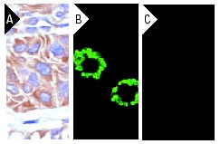
Cox-2 (M-19): sc-1747. Immunoperoxidase staining of formalin-fixed, paraffin-embedded human lung tumor showing membrane and cytoplasmic staining (A). Immunofluorescence staining of methanol-fixed RAW 264.7 cells induced with LPS and PMA showing cytoplasmic vesicle localization (B) and untreated control RAW 264.7 cells (C).
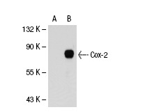
Cox-2 (M-19): sc-1747. Western blot analysis of Cox-2 expression in non-transfected: sc-110760 (A) and human Cox-2 transfected: sc-113099 (B) 293 whole cell lysates.
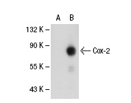
Cox-2 (M-19)-R: sc-1747-R. Western blot analysis of Cox-2 expression in non-transfected: sc-110760 (A) and human Cox-2 transfected: sc-113099 (B) 293 whole cell lysates.
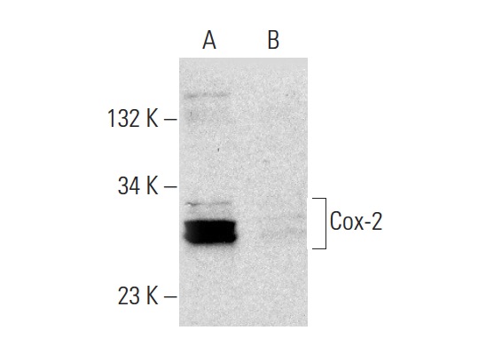
Cox-2 (M-19): sc-1747. Western blot analysis of Cox-2 expression in untreated (A) and DuP-697 (sc-200680) treated (B) A549 whole cell lysates. Note down regulation of Cox-2 expression in lane B.
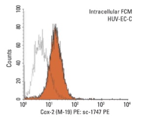
Cox-2 (M-19) PE: sc-1747 PE. Intracellular FCM analysis of fixed and permeabilized HUV-EC-C cells. Black line histogram represents the isotype control, normal goat IgG: sc-3992.


