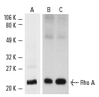
Western blot analysis of Rho A expression in A-431 (A), HeLa (B) and KNRK (C) whole cell lysates. Antibodies tested include Rho A (119): sc-179 (A) and Rho A (26C4): sc-418 (B,C).
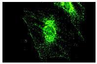
Rho A (119): sc-179. Cytoplasmic immunofluorescence staining of methanol/acetone-fixed rat embryo fibroblasts using fluorescein-labeled goat anti-rabbit IgG secondary antibody.
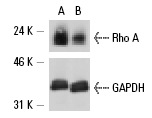
Rho A siRNA (h,m): sc-29471. Western blot analysis of Rho A expression in non-transfected control (A) and Rho A siRNA (B) transfected HeLa cells. Blot probed with Rho A (119): sc-179. GAPDH (V-18): sc-20357 used as specificity and loading control.
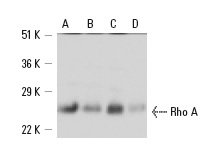
Rho A (119): sc-179. Western blot analysis of Rho A expression in KNRK (A), PC-12 (B), HL-60 (C) and MCF7 (D) whole cell lysates.
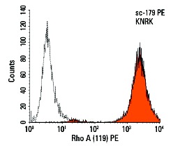
Rho A (119) PE: sc-179 PE. Intracellular FCM analysis of fixed and permeabilized KNRK cells. Black line histogram represents the isotype control, normal rabbit IgG: sc-3871.
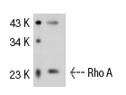
Rho A (119): sc-179. Western blot analysis of Rho A expression in Jurkat whole cell lysate.
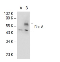
Rho A (119)-G: sc-179-G. Western blot analysis of Rho A expression in non-transfected: sc-117752 (A) and human Rho A transfected: sc-177860 (B) 293T whole cell lysates.
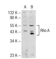
Rho A (119): sc-179. Western blot analysis of Rho A expression in non-transfected: sc-117752 (A) and human Rho A transfected: sc-177860 (B) 293T whole cell lysates.
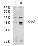
Rho A (119): sc-179. Western blot analysis of Rho A expression in non-transfected: sc-117752 (A) and human Rho A transfected: sc-177861 (B) 293T whole cell lysates.
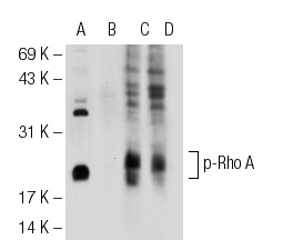
Western blot analysis of Rho A phosphorylation in untreated (A,C) and lambda protein phosphatase treated (B,D) mouse brain tissue extracts. Antibodies tested include p-Rho A (Ser 188): sc-32954 (A,B) and Rho A (119): sc-179 (C,D).
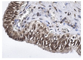
Rho A (119): sc-179. Immunoperoxidase staining of formalin fixed, paraffin-embedded human testis tissue showing nuclear and cytoplasmic staining of cells in seminiferous ducts.


