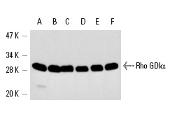
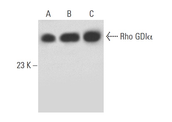
Rho GDIα (A-20): sc-360. Western blot analysis of Rho GDIα expression in KNRK (A), PC-12 (B), HL-60 (C), HeLa (D), SK-BR-3 (E) and MCF7 (F) whole cell lysates.
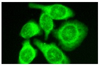
Rho GDIα (A-20): sc-360. Immunofluorescence staining of methanol-fixed HeLa cells showing cytoplasmic staining.
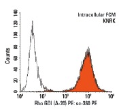
Rho GDIα (A-20) PE: sc-360 PE. Intracellular FCM analysis of fixed and permeabilized KNRK cells. Black line histogram represents the isotype control, normal rabbit IgG: sc-3871.
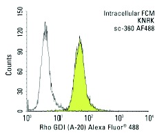
Rho GDIα (A-20) Alexa Fluor 488 : sc-360 AF488. Intracellular FCM analysis of fixed and permeabilized KNRK cells. Black line histogram represents the isotype control, normal rabbit IgG: sc-45068.
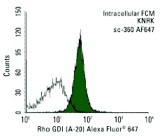
Rho GDIα (A-20) Alexa Fluor 647 : sc-360 AF647. Intracellular FCM analysis of fixed and permeabilized KNRK cells. Black line histogram represents the isotype control, normal rabbit IgG: sc-24647.
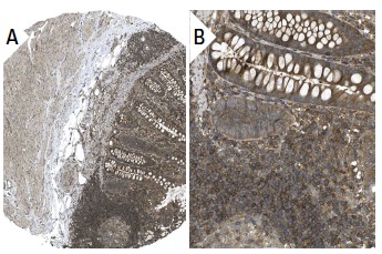
Rho GDIaα (A-20): sc-360. Immunoperoxidase staining of formalin fixed, paraffin-embedded human appendix tissue showing cytoplasmic staining of lymphoid and glandular cells at low (A) and high (B) magnification. Kindly provided by The Swedish Human Protein Atlas (HPA) program.
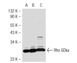
Rho GDIα (A-20): sc-360. Western blot analysis of Rho GDIα expression in non-transfected 293T: sc-117752 (A), mouse Rho GDIα transfected 293T: sc-125905 (B) and K-562 (C) whole cell lysates.
Rho GDIα (A-20): sc-360. Western blot analysis of Rho GDIα expression in non-transfected 293T: sc-117752 (A), human Rho GDIα transfected 293T: sc-111739 (B) and MCF7 (C) whole cell lysates.
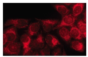
Rho GDIα (A-20): sc-360. Immunofluorescence staining of methanol-fixed HeLa cells showing cytoplasmic localization.