Western blot analysis of extracts from human fibroblasts synchronized by serum deprivation, using Phospho-Rb (Ser795) Antibody. Cells were synchronized for 24 hours, then released by addition of serum and harvested at the times indicated. Cell cycle progression was verified by cyclin analysis and FACS. (Provided by John Boylan, Dupont/Merck, Delaware.)

Western blot analysis of Rb-C Fusion Protein #6022 (amino acids 701-928 of Rb fused to MBP) before (-) and after (+) in vitro phosphorylation by cdc2/cyclin B Protein Kinase (New England Biolabs #P6020), using Phospho-Rb (Ser795) Antibody #9301 (left) or the control antibody (right).

Western blot analysis of extracts from human fibroblasts synchronized by serum deprivation, using Phospho-Rb (Ser780) Antibody. Cells were synchronized for 24 hours then released by addition of serum and harvested at the times indicated. Cell cycle progression was verified by cyclin analysis and FACS. (Provided by John Boylan, Dupont/Merck, Delaware.)

Western blot analysis of extracts from human fibroblasts synchronized by serum deprivation, using Phospho-Rb (Ser807/811) Antibody. Cells were synchronized for 24 hours then released by addition of serum and harvested at the times indicated. Cell cycle progression was verified by cyclin analysis and FACS. (Provided by John Boylan, Dupont/Merck, Delaware.)

Western blot analysis of extracts from HeLa cells, transfected with 100 nM SignalSilence ® Control siRNA (Fluorescein Conjugate) #6201 (-), SignalSilence ® Rb siRNA I #6451 (+) or SignalSilence ® Rb siRNA II (+), using Rb (4H1) Mouse mAb #9309 and α-Tubulin (11H10) Rabbit mAb #2125. The Rb (4H1) Mouse mAb confirms silencing of Rb expression, while the α-Tubulin (11H10) Rabbit mAb is used to control for loading and specificity of Rb siRNA.

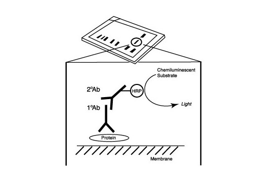
After the primary antibody is bound to the target protein, a complex with HRP-linked secondary antibody is formed. The LumiGLO ® is added and emits light during enzyme catalyzed decomposition.

Western blot analysis of Rb Control Protein #9303, using Phospho-Rb (Ser795) Antibody (upper) or Rb (4H1) mAb #9309 (lower).

Western blot analysis of extracts from COS-7 cells, untreated or hydroxyurea-treated (G1/S), using Rb (4H1) Mouse mAb.

Immunohistochemical analysis of paraffin-embedded human breast carcinoma, showing nuclear localization, using Rb (4H1) Mouse mAb.

Immunohistochemical analysis of paraffin-embedded human lung carcinoma, using Rb (4H1) Mouse mAb.

Flow cytometric analysis of Jurkat cells, using Rb (4H1) Mouse mAb versus propidium iodide (DNA content). The box indicates Rb positive cells.

Confocal immunofluorescent image of SH-SY5Y cells, using RB (4H1) Mouse mAb (green). Actin filaments have been labeled with Alexa Fluor® 555 phalloidin (red).

Chromatin immunoprecipitations were performed with cross-linked chromatin from 4 x 10 6 Raji cells and either 5 μl of Rb (4H1) Mouse mAb or 2 μl of Normal Rabbit IgG #2729 using SimpleChIP ® Enzymatic Chromatin IP Kit (Magnetic Beads) #9003. The enriched DNA was quantified by real-time PCR using using SimpleChIP ® Human Timeless Intron 1 Primers #7001, human DHFR promoter primers, and SimpleChIP ® Human α Satellite Repeat Primers #4486. The amount of immunoprecipitated DNA in each sample is represented as signal relative to the total amount of input chromatin, which is equivalent to one.


