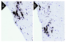
CD68 (KP1): sc-20060. Immunoperoxidase staining of formalin-fixed paraffin embedded human coronary artery tissue showing macrophage staining adjacent to the lumina (A,B). Kindly provided by Dr. John Sanders, University of Virginia.
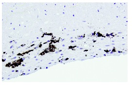
CD68 (KP1): sc-20060. Immunoperoxidase staining of formalin-fixed paraffin embedded human coronary artery tissue showing macrophage staining adjacent to the lumina (A,B). Kindly provided by Dr. John Sanders, University of Virginia.
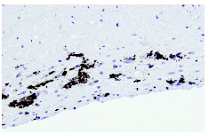
CD68 (KP1): sc-20060. Immunoperoxidase staining of formalin-fixed paraffin embedded human coronary artery tissue showing macrophage staining adjacent to the lumina (A,B). Kindly provided by Dr. John Sanders, University of Virginia.
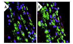
CD68 (KP1): sc-20060. Immunofluorescence staining of formalin-fixed paraffin embedded human coronary artery tissue showing CD68 staining of macrophages (green), DAPI counterstain (blue) and tissue autofluorescence (red) (A,B). Kindly provided by Dr. John Sanders, University of Virginia.
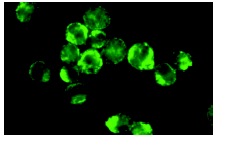
CD68 (KP1): sc-20060. Immunofluorescence staining of methanol-fixed THP-1 cells showing cytoplasmic and membrane localization.
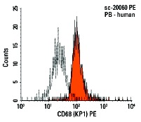
CD68 (KP1) PE: sc-20060 PE. FCM analysis of human peripheral blood leukocytes. Black line histogram represents the isotype control, normal mouse IgG
1: sc-2866.
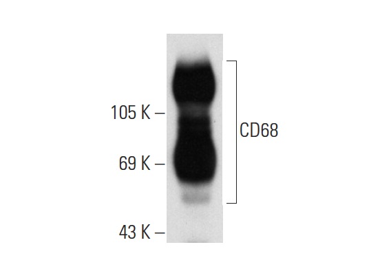
CD68 (KP1): sc-20060. Western blot analysis of CD68 expression in THP-1 whole cell lysate.
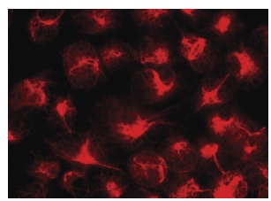
CD68 (KP1): sc-20060. Immunofluorescence staining of methanol-fixed HeLa cells showing membrane localization.
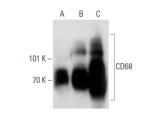
CD68 (KP1): sc-20060. Western blot analysis of CD68 expression in K-562 (A), U-937 (B) and AML-193 (C) whole cell lysates.
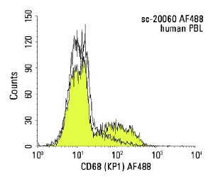
CD68 (KP1) AF488: sc-20060 AF488. FCM analysis of human peripheral blood leukocytes. Black line histogram represents the isotype control, normal mouse IgG
1: sc-3890.
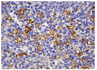
CD68 (KP1): sc-20060. Immunoperoxidase staining of formalin fixed, paraffin-embedded human spleen tissue showing cytoplasmic staining of subset of cells in red pulp.


