 Immunofluorescence
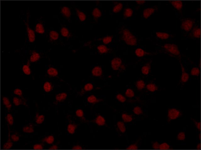
Predominant nuclear localization of TNPO2 in HEK-293T cells. Cells were fixed and stained with 5 μg/mL Rabbit Anti-TNPO2 (N-terminal) (
Cat. No. SAB4200032) followed by Goat Anti-Rabbit, Cy3 conjugated (×40 magnification).
 Immunoblotting
Lysates of various cell lines were separated on SDS-PAGE and probed with 4 μg/mL Rabbit Anti-TNPO2 (N-terminal) (
Cat. No. SAB4200032). The antibody was developed with Goat Anti-Rabbit IgG (
Cat. No. A0545) Peroxidase conjugate and a chemiluminescence substrate.
Lanes1. HEK-293T cells; 2. Jurkat; 3. HeLa; 4. K562; 5. MCF7
 Immunoprecipitation
Rabbit Anti-TNPO2 (N-terminal) (
Cat. No. SAB4200031) and Rabbit Anti-TNPO2 (C-terminal) (
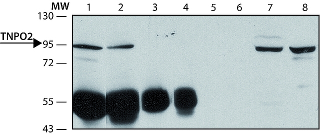
Cat. No. SAB4200032) were used to immunoprecipitate human TNPO2 from lysate of HEK-293T cells over-expressing human TNPO2.
Lanes1. IP antibody: 10 μg Anti-TNPO2 (N-terminal) (
Cat. No. SAB4200032)
2. IP antibody: 10 μg Anti-TNPO2 (C-terminal) (
Cat. No. SAB4200031)
3. Negative control: Anti-TNPO2 (N-terminal) IP without cell lysate
4. Negative control: Anti-TNPO2 (C-terminal) IP without cell lysate
5. Negative control: Anti-TNPO2 (N-terminal) IP without IP antibody
6. Negative control: Anti-TNPO2 (C-terminal) IP without IP antibody
7. Immunoblotting positive control: total HEK-293T cells over-expressing human TNPO2 blot with Anti-TNPO2 (N-terminal) (
Cat. No. SAB4200032)
8. Immunoblotting positive control: total HEK-293T cells over-expressing human TNPO2 blot with Anti-TNPO2 (C-terminal) (


