 Immunofluorescence
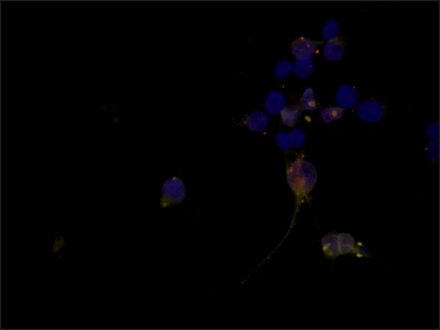
Cytoplasmic P-bodies localization of eIF2C1/Ago1 in 293T cells over-expressing FLAG-tagged eIF2C1/Ago1. Cells were fixed and co-stained with 5 μg/mL Rabbit Anti-eIF2C1/Ago1 (C-terminal) (
Cat. No. SAB4200065) (Red) and 1:1,000 Anti-FLAG (
Cat. No. F3165) (Green) followed by Goat Anti-Rabbit, Cy3 conjugated and Goat Anti-Mouse IgG, Atto 488 conjugated (
Cat. No. 62197) respectively. The cells were counterstained with DAPI (Blue) to stain nuclei (×40 magnification).
 Immunoblotting
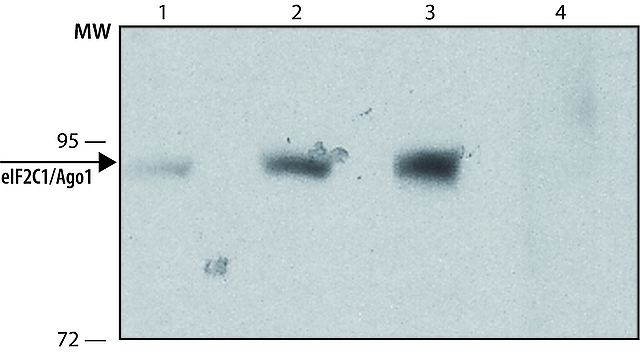
Lysate of HEK-293T cells over-expressing human eIF2C1/Ago1 was separated on SDS-PAGE and probed with 1-4 μg/mL Anti-eIF2C1/Ago1 (
Cat. No. SAB4200065). The antibody was developed with Goat Anti-Rabbit IgG, Peroxidase conjugate (
Cat. No. A0545) and a chemiluminescence substrate.
Lanes1. Antibody 1 μg/mL
2. Antibody 2 μg/mL
3. Antibody 4 μg/mL
4. Antibody 4 μg/mL + 20 μg/mL Anti-eIF2C1/Ago1 immunizing peptide
 Immunoprecipitation
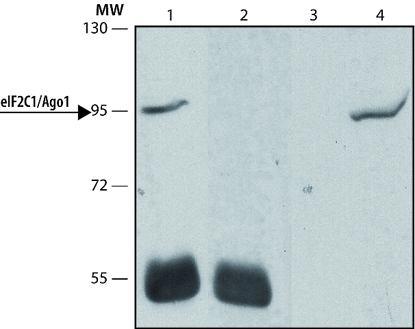
Rabbit Anti-eIF2C1/Ago1 (
Cat. No. SAB4200065) was used to immunoprecipitate human eIF2C1/Ago1 from lysate of HEK-293T cells over-expressing human eIF2C1 /Ago1. Detection antibody: 2 μg/mL Anti-anti-eIF2C1/Ago1 (N-terminal) (
Cat. No. SAB4200066).
Lanes 1. IP antibody: 10 μg
2. Negative control: without cell lysate
3. Negative control: without IP antibody
4. Immunobloting positive control: total HEK-293T cells over-expressing human eIF2C1/Ago1. Detection antibody: 4 μg/mL Anti-eIF2C1/Ago1 (
Cat. No. SAB4200065)


