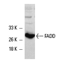
FADD (H-181): sc-5559. Western blot analysis of human recombinant FADD expression in A-431 cells.
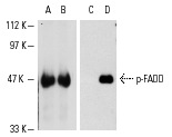
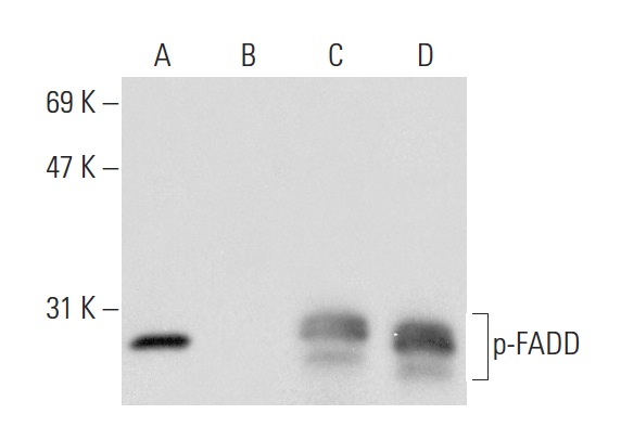
Western blot analysis of human recombinant FADD (A,C) and FADD phosphorylated by ERK 2 (B,D). Antibodies tested include FADD (H-181): sc-5559 (A,B) and p-FADD (Ser 194): sc-12439 (C,D).
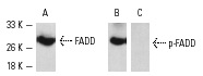
Western blot detection of FADD phosphorylation using A-431 cells. Blots were probed with FADD (H-181): sc-5559 (A) and p-FADD (Ser 194)-R: sc-12439-R (B,C). In B and C, the antibody was preincubated with cognate non-phosphorylated or phosphorylated peptide, respectively.

FADD (H-181): sc-5559. Western blot analysis of FADD expression in non-transfected: sc-117752 (A) and mouse FADD transfected: sc-126821 (B) 293T whole cell lysates.
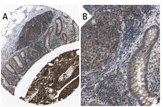
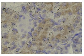
FADD (H-181): sc-5559. Immunoperoxidase staining of formalin fixed, paraffin-embedded human appendix tissue showing cytoplasmic staining of glandular and lymphoid cells (low and high magnification). Kindly provided by The Swedish Human Protein Atlas (HPA) program.
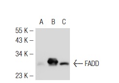
FADD (H-181): sc-5559. Western blot analysis of FADD expression in non-transfected 293T: sc-117752 (A), mouse FADD transfected 293T: sc-126822 (B) and SW480 (C) whole cell lysates.
Western blot analysis of FADD phosphorylation in untreated (A,C) and treated (B,D) A-431 whole cell lysates. Antibodies tested include p-FADD (Ser 194): sc-12439 (A,B) and FADD (H-181): sc-5559 (C,D).
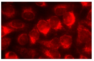
FADD (H-181): sc-5559. Immunofluorescence staining of methanol-fixed HeLa cells showing cytoplasmic localization.
FADD (H-181): sc-5559. Immunoperoxidase staining of formalin fixed, paraffin-embedded mouse kidney tissue showing cytoplasmic localization.