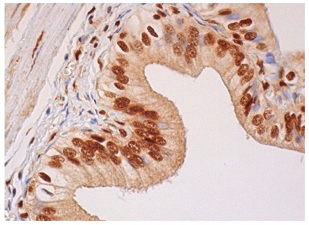
p-FADD (Ser 194): sc-12439. Immunoperoxidase staining of formalin fixed, paraffin-embedded human gall bladder tissue showing nuclear staining of glandular cells.
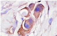
p-FADD (Ser 194): sc-12439. Immunoperoxidase staining of formalin-fixed, paraffin-embedded human breast tumor showing cytoplasmic staining.
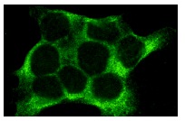
p-FADD (Ser 194): sc-12439. Immunofluorescence staining of methanol-fixed A-431 cells showing cytoplasmic staining.
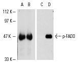
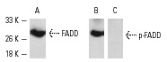
Western blot analysis of human recombinant FADD (A,C) and FADD phosphorylated by ERK 2 (B,D). Antibodies tested include FADD (H-181): sc-5559 (A,B) and p-FADD (Ser 194): sc-12439 (C,D).
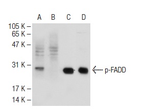
Western blot detection of FADD phosphorylation using A-431 cells. Blots were probed with FADD (H-181): sc-5559 (A) and p-FADD (Ser 194)-R: sc-12439-R (B,C). In B and C, the antibody was preincubated with cognate non-phosphorylated or phosphorylated peptide, respectively.
Western blot analysis of FADD phosphorylation in untreated (A,C), and lambda protein phosphatase treated (B,D) A-431 whole cell lysates. Antibodies tested include p-FADD (Ser 194): sc-12439 (A,B) and FADD (FD19): sc-56093 (B,D).
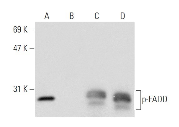
Western blot analysis of FADD phosphorylation in untreated (A,C) and treated (B,D) A-431 whole cell lysates. Antibodies tested include p-FADD (Ser 194): sc-12439 (A,B) and FADD (H-181): sc-5559 (C,D).
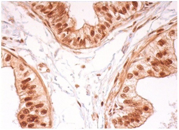
p-FADD (Ser 194)-R: sc-12439-R. Immunoperoxidase staining of formalin fixed, paraffin-embedded human epididymis tissue showing nuclear staining of glandular cells.