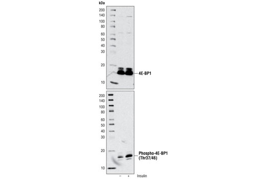
Western blot analysis of extracts from 293T cells using 4E-BP1 Antibody #9452 (upper) and Phospho-4E-BP1 (Thr37/46) Antibody #2855 (lower). The cells were starved for 24 hours in serum-free medium and underwent a 1 hour amino acid deprivation. Amino acids were replenished for 1 hour. Cells were then either untreated (-) or treated with 100 nM insulin (+) for 30 minutes.

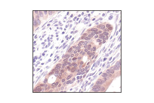
Immunohistochemical analysis of paraffin-embedded human colon carcinoma using Phospho-4E-BP1 (Thr37/46) (236B4) Rabbit mAb.

Immunohistochemical analysis of paraffin-embedded human lymphoma using Phospho-4E-BP1 (Thr37/46) (236B4) Rabbit mAb.

Immunohistochemical analysis using Phospho-4E-BP1 (Thr37/46) (236B4) Rabbit mAb on SignalSlide (TM) Phospho-Akt (Ser473) IHC Controls #8101 (paraffin-embedded LNCaP cells untreated (left) or LY294002-treated (right)).

Immunohistochemical analysis of paraffin-embedded human colon carcinoma using Phospho-4E-BP1 (Thr37/46) (236B4) Rabbit mAb in the presence of control peptide (left) or Phospho-4E-BP1 (Thr37/46) Blocking Peptide #1052 (right).

Confocal immunofluorescent analysis of 293 cells, expressing either non-targeting shRNA (top) or shRNA targeting 4E-BP1/2 (bottom), using Phospho-4E-BP1 (Thr37/46) (236B4) Rabbit mAb (green). To confirm phospho-specificity, cells were treated with an inhibitor cocktail consisting of LY294002 #9901, U0126 #9903, and Rapamycin #9904 (50 μM; 10 μm; 10 nM; 2 hr) (left), stimulated with insulin (100 nM, 30 min; middle), or processed with λ-phosphatase following insulin stimulation (right). Red = Propidium Iodide (PI)/RNase Staining Solution (#4087).

Flow cytometric analysis of Jurkat cells, untreated (green) or LY294002, Wortmannin and U0126-treated (blue), using Phospho-4E-BP1 (Thr36/46) (236B4) Rabbit mAb compared to a nonspecific negative control antibody (red).


