 Immunohistochemistry
Anti-TRIT1:
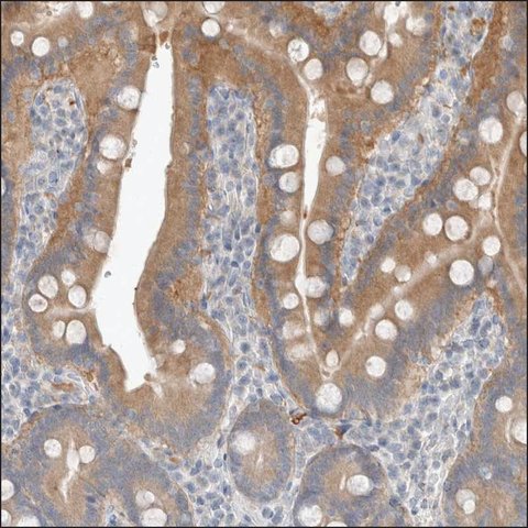
Cat. No. HPA024174: Immunohistochemical staining of human duodenum shows moderate cytoplasmic positivity in glandular cells.
 Immunofluorescence
Anti-TRIT1:
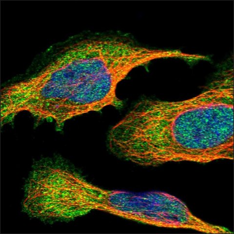
Cat. No. HPA024174: Immunofluorescent staining of Human cell line U-2 OS shows positivity in nucleus but not nucleoli, plasma membrane and cytoplasm.
![<B>Immunoblotting</B><BR/>Anti-TRIT1: <B>Cat. No. HPA024174</B>: <BR/><B>Lanes</B><BR/>1. Marker [kDa] 230, 130, 95, 72, 56, 36, 28, 17, 11<BR/>2. Human cell line RT-4](http://www.bioprodhub.com/system/product_images/ab_products/7/sub_4/3808_hpa024174-blot-1-l-large.jpg) Immunoblotting
ImmunoblottingAnti-TRIT1:
Cat. No. HPA024174:
Lanes1. Marker [kDa] 230, 130, 95, 72, 56, 36, 28, 17, 11
2. Human cell line RT-4
 Immunoblotting
Anti-TRIT1:
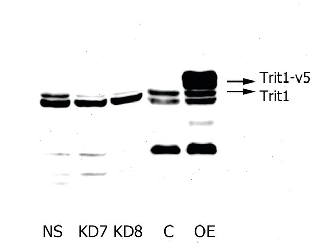
Cat. No. HPA024174: Western-blot to test TRIT1 expression in HepG2 cells. RIPA lysates (50 μg) from two different HepG2 knockdown cell lines (KD7 and KD8), from overexpressing v5-tagged TRIT1 HepG2 cells (OE) and lysates from their respective controls (NS and C) were separated by SDS-PAGE electrophoresis. Transferred nitrocellulose membranes were subjected to immunoblotting with Rabbit polyclonal Anti-TRIT1 (Sigma 1:1,000 dilution). Luminiscence was detected by FluorChem F2 camera using 1:2,000 HRP conjugated Anti-Rabbit IG antibody (Dako).
Submitted by Dr N. Fradejas, Charité-Universitätsmedizin, Berlin.


![<B>Immunoblotting</B><BR/>Anti-TRIT1: <B>Cat. No. HPA024174</B>: <BR/><B>Lanes</B><BR/>1. Marker [kDa] 230, 130, 95, 72, 56, 36, 28, 17, 11<BR/>2. Human cell line RT-4](http://www.bioprodhub.com/system/product_images/ab_products/7/sub_4/3808_hpa024174-blot-1-l-large.jpg)
