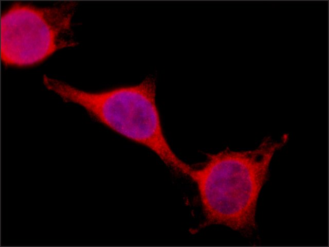
 Immunofluorescence
Staining of PSPC1 in HEK-293T cells. Cells were fixed and stained with 1.25 μg/mL Rabbit Anti-PSPC1 (C-Terminal) (
Cat. No. SAB4200067) followed by Goat Anti-Rabbit, Cy3 conjugate and counterstained with DAPI (blue) to stain nuclei (×40 magnification).
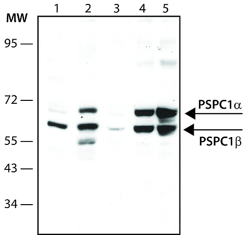
 Immunoblotting
Lysates of the indicated cells were separated on SDS-PAGE and probed with 2 μg/mL Rabbit Anti-PSPC1 (C-Terminal) (
Cat. No. SAB4200067). The antibody was developed with Goat Anti-Rabbit IgG, Peroxidase conjugate (
Cat. No. A0545) and a chemiluminescence substrate.
Lanes1. HeLa; 2. K-562; 3. Raw264; 4. HEK-293T; 5. HEK-293T over-expressing PSPC1
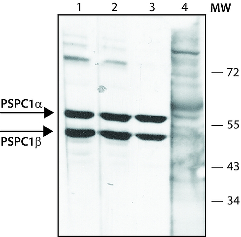
 Immunoblotting
Lysate of HEK-293Tcells was separated on SDS-PAGE and probed with Rabbit Anti-PSPC1 (C-terminal) (
Cat. No. SAB4200067). The antibody was developed with Goat Anti-Rabbit IgG, Peroxidase conjugate (
Cat. No. A0545) and a chemiluminescence substrate.
Lanes1. Antibody 2 μg/mL
2. Antibody 1 μg/ mL
3. Antibody 0.5 μg/ mL
4. Antibody 1 μg/mL + 10 μg/mL PSPC1 immunizing peptide
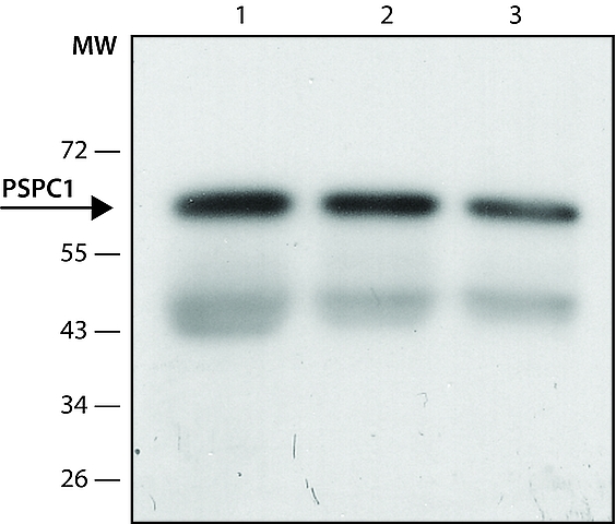
 Immunoprecipitation
Rabbit Anti-PSPC1 (C-Terminal) (
Cat. No. SAB4200067) was used to immunoprecipitate human PSPC1 from lysate of HEK-293T cells. Detection Antibody: Rabbit Anti-PSPC1 (N-Terminal) (
Cat. No. SAB4200068).
Lanes1. IP antibody: 10 μg
2. IP antibody: 5 μg
3. IP antibody: 2.5 μg


