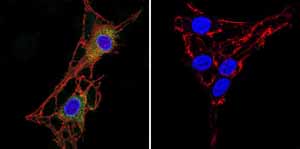
Immunofluorescent analysis of Acetylcholinesterase using Acetylcholinesterase Monoclonal antibody (ZR3) ab2802 shows staining in C6 glioma cells. Acetylcholinesterase staining (green) F-Actin staining with Phalloidin (red) and nuclei with DAPI (blue) is shown. Cells were grown on chamber slides and fixed with formaldehyde prior to staining. Cells were probed without (control) or with or an antibody recognizing Acetylcholinesterase ab2802 at a dilution of 1:20 over night at 4 ?C washed with PBS and incubated with a DyLight-488 conjugated secondary antibody. Images were taken at 60X magnification.

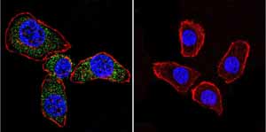
Immunofluorescent analysis of Acetylcholinesterase using Acetylcholinesterase Monoclonal antibody (ZR3) ab2802 shows staining in U251 glioma cells. Acetylcholinesterase staining (green) F-Actin staining with Phalloidin (red) and nuclei with DAPI (blue) is shown. Cells were grown on chamber slides and fixed with formaldehyde prior to staining. Cells were probed without (control) or with or an antibody recognizing Acetylcholinesterase ab2802 at a dilution of 1:20 over night at 4 ?C washed with PBS and incubated with a DyLight-488 conjugated secondary antibody. Images were taken at 60X magnification.

