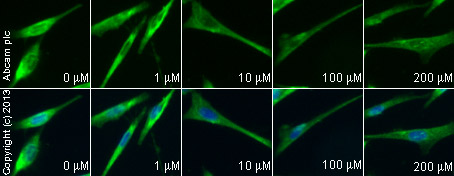
ab13847 staining caspase 3 in SKNSH cells treated with Z-IETD-FMK (ab141382), by ICC/IF. Decrease in caspase 3 expression correlates with increased concentration of Z-IETD-FMK, as described in literature.The cells were incubated at 37°C for 1 hour in media containing different concentrations of ab141382 (Z-IETD-FMK) in DMSO. After this incubation, 10 μM of camptothecin (ab120115) was added to all samples and the cells were incubated for further 24 hours. The samples were then fixed with 4% formaldehyde for 10 minutes at room temperature and blocked with PBS containing 10% goat serum, 0.3 M glycine, 1% BSA and 0.1% tween for 2h at room temperature. Staining of the treated cells with ab13847 (5 μg/ml) was performed overnight at 4°C in PBS containing 1% BSA and 0.1% tween. A goat anti-rabbit DyLight 488 (ab96899) at 1/250 dilution was used as the secondary antibody. Nuclei were counterstained with DAPI and are shown in blue.
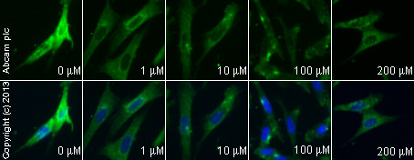
ab13847 staining caspase 3 in SKNSH cells treated with Z-VDVAD-FMK (ab142035), by ICC/IF. Decrease in caspase 3 expression correlates with increased concentration of Z-VDVAD-FMK, as described in literature.The cells were incubated at 37°C for 1 hour in media containing different concentrations of ab142035 (Z-VDVAD-FMK) in DMSO. After this incubation, 10 μM of camptothecin (ab120115) was added to all samples and the cells were incubated for further 24 hours. The samples were then fixed with 4% formaldehyde for 10 minutes at room temperature and blocked with PBS containing 10% goat serum, 0.3 M glycine, 1% BSA and 0.1% tween for 2h at room temperature. Staining of the treated cells with ab13847 (5 μg/ml) was performed overnight at 4°C in PBS containing 1% BSA and 0.1% tween. A anti-rabbit DyLight 488 (ab96899) at 1/250 dilution was used as the secondary antibody. Nuclei were counterstained with DAPI and are shown in blue.
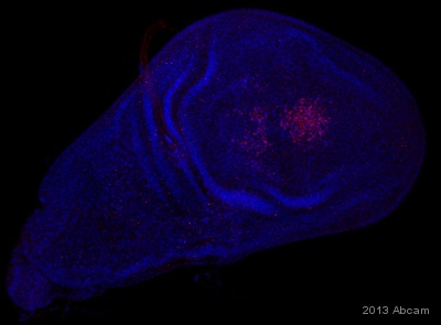
ab13847 staining Drosophila melanogaster 3rd instar wing imaginal disc (red) by IHC-wholemount. The tissue was fixed with 4% formaldehyde in PBS at room temperature for 20 minutes. Sample was blocked in PBS+ Triton-X 100 0.3% + BSA 1% for 1h at room temperature, followed by staining with ab13847 at 1/500 (in PBS+ Triton-X 100 0.3% + BSA 1%) for 16h at 4°C. A Cy3 conjugated goat anti-rabbit polyclonal secondary antibody at 1/200 was incubated for 2h at room temperature. Nuclei were stained with DAPI.See Abreview
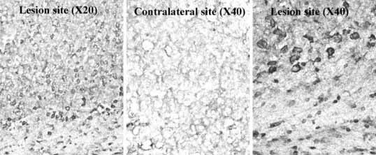
ab13847 was used in IHC of frozen sections from a rat brain with a kainite lesion. The non lesionned contralateral site serves as a negative control. The sections were fixed with paraformaldehyde. The tissue was perfused with 4% PFA and embedded in OCT compound and cut on the cryostat. The primary antibody was incubated for 12 hours at a dilution of 1/300. A biotin labelled secondary antibody was used at a dilution of 1/300.
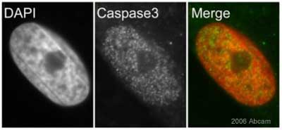
ab13847 at a dilution of 1/500 staining Asynchronous HeLa cells by Immunocytochemistry. The antibody was incubated with the cells for 30 minutes and then detected using a Cy3 conjugated Goat Anti-Rabbit IgG (H+L). This antibody gives a predominantly nuclear focal staining pattern in all interphase nuclei investigated.This image is courtesy of an Abreview by Kirk McManus submitted on 2 December 2005.See Abreview
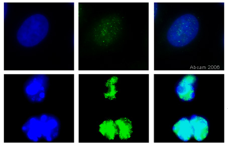
HeLa cells were fixed for 10 minutes at room temperature in 3.7% PFA and permeabilised in 0.1% Triton X-100/PBS then incubated with ab13847 (5µg/ml) for 1 hour at room temperature. The top panel shows control cells treated with DMSO. The bottom panel shows HeLa cells treated with 1 mM staurosporine for 4 hours to induce caspase-3 activation. ab13847 staining is shown in green and counterstaining with DAPI is shown in blue. 100x magnification.The image shows the staining with ab13847 is very faint in the untreated control cultures, but very bright after activation of capsase-3 by treatment with the staurosporine. (N.B. in these cultures the nuclei are apoptotic).
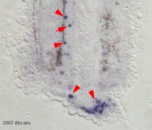
ab13847 at 1/1000 staining Xenopus laevis wholemount and sectioned tails by IHC-P. The tissue was formaldehyde fixed and blocked prior to incubation with the antibody for 12 hours. An alkaline phosphatase conjugated goat anti-rabbit antibody was used as the secondary.See Abreview
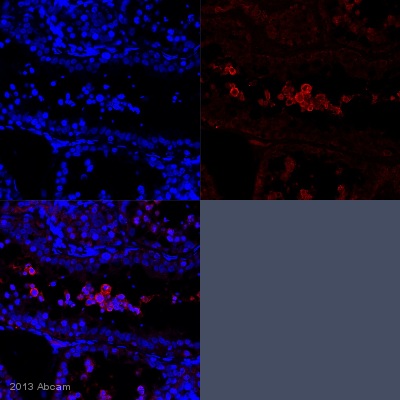
IHC-P image of Caspase 3 staining with ab13847 on tissue sections from juvenile marmoset testis. The sections were subjected to heat-mediated antigen retrieval using Dako antigen retrieval solution. The sections were then blocked with 5% milk for 30 minutes at 25°C, before incubation with ab13847 (1/100 dilution) for 18 hours at 4°C. The secondary was an Alexa-Fluor 555 conjugated goat anti-rabbit polyclonal, used at a 1/500 dilution.See Abreview
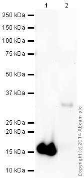
All lanes : Anti-active + pro Caspase 3 antibody (ab13847) at 1 µg/mlLane 1 : Human Caspase 3 (active) Recombinant ProteinLane 2 : Human Pro Caspase 3 (inactive) Recombinant ProteinLysates/proteins at 0.1 µg per lane.SecondaryGoat Anti-Rabbit IgG H&L (HRP) (ab97051) at 1/10000 dilutiondeveloped using the ECL techniquePerformed under reducing conditions.
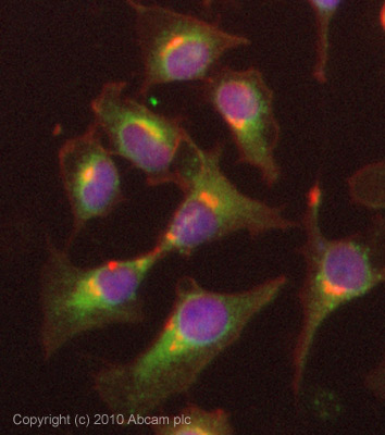
ICC/IF image of ab13847 stained HeLa cells. The cells were 4% PFA fixed (10 min) and then incubated in 1%BSA / 10% normal goat serum / 0.3M glycine in 0.1% PBS-Tween for 1h to permeabilise the cells and block non-specific protein-protein interactions. The cells were then incubated with the antibody (ab13847, 5µg/ml) overnight at +4°C. The secondary antibody (green) was Alexa Fluor® 488 goat anti-rabbit IgG (H+L) used at a 1/1000 dilution for 1h. Alexa Fluor® 594 WGA was used to label plasma membranes (red) at a 1/200 dilution for 1h. DAPI was used to stain the cell nuclei (blue) at a concentration of 1.43µM. This antibody also gave a positive result in 4% PFA fixed (10 min) Hek293, HepG2 and MCF7 cells at 5µg/ml, and in 100% methanol fixed (5 min) HeLa, Hek293 and MCF7 cells at 5µg/ml.
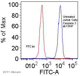
ab13847 staining active caspase 3 in Human Jurkat cells by Flow Cytometry. Cells were prepared in a phosphate buffered solution containing 0.1% sodium azide with FBS fixed with paraformaldehyde and permeabilized with Triton X-100 and NP40. The sample was incubated with the primary antibody (1/100 in wash buffer) for 24 hours at 4°C. A FITC-conjugated Goat anti-rabbit Ig (1/100) was used as the secondary antibody.Gating Strategy: Isolate cell population from plot of SSC-A / FSA-A
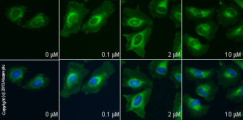
ab13847 staining caspase 3 in HeLa cells treated with RTIL-13™ (ab120465), by ICC/IF. Increase in caspase 3 expression correlates with increased concentration of RTIL-13™, as described in literature.The cells were incubated at 37°C for 24h in media containing different concentrations of ab120465 (RTIL-13™) in DMSO, fixed with 4% formaldehyde for 10 minutes at room temperature and blocked with PBS containing 10% goat serum, 0.3 M glycine, 1% BSA and 0.1% tween for 2h at room temperature. Staining of the treated cells with ab13847 (10 µg/ml) was performed overnight at 4°C in PBS containing 1% BSA and 0.1% tween. A goat anti-rabbit DyLight 488 secondary antibody (ab96899) at 1/250 dilution was used as the secondary antibody. Nuclei were counterstained with DAPI and are shown in blue.

ab13847 staining caspase 3 in A549 cells treated with quercetin (ab120247), by ICC/IF. Increase in caspase 3 expression correlates with increased concentration of quercetin, as described in literature.The cells were incubated at 37°C for 6h in media containing different concentrations of ab120247 (quercetin) in DMSO, fixed with 100% methanol for 5 minutes at -20°C and blocked with PBS containing 10% goat serum, 0.3 M glycine, 1% BSA and 0.1% tween for 2h at room temperature. Staining of the treated cells with ab13847 (1 µg/ml) was performed overnight at 4°C in PBS containing 1% BSA and 0.1% tween. A goat anti-rabbit DyLight 488 polyclonal secondary antibody (ab968999) at 1/250 dilution was used as the secondary antibody. Nuclei were counterstained with DAPI and are shown in blue.


