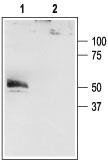
Western blot analysis of HEK-293-K2P4.1 transfected cells: 1. Anti-K2P4.1 (TRAAK) antibody (#AG1103), (1:200). 2. Anti-K2P4.1 (TRAAK) antibody, preincubated with the control peptide antigen.

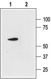
Western blot analysis of rat cerebellum lysate:

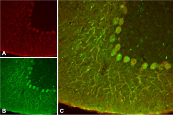
Expression of K2P4.1 in rat cerebellum Immunohistochemical staining of rat cerebellum using Anti- K2P4.1 (TRRAK) antibody (#AG1103 ). A. K2P4.1 channel appears in Purkinje neuronal processes (red). B. Staining of Purkinje nerve cells with mouse anti-calbindin D28K (a calcium binding protein, green). C. Confocal merge of K2P4.1 channel and calbindin D28K demonstrates the co-localization of these proteins.


