![All lanes : Anti-ADAR1 antibody [EPR7033] (ab126745) at 1/1000 dilutionLane 1 : HeLa (treated with IFN-alpha) cell lysateLane 2 : HeLa cell lysateLane 3 : Ramos cell lysateLane 4 : SH-SY5Y cell lysateLysates/proteins at 10 µg per lane.SecondaryHRP labelled goat anti-rabbit at 1/2000 dilution](http://www.bioprodhub.com/system/product_images/ab_products/2/sub_1/2824_ADAR1-Primary-antibodies-ab126745-1.jpg)
All lanes : Anti-ADAR1 antibody [EPR7033] (ab126745) at 1/1000 dilutionLane 1 : HeLa (treated with IFN-alpha) cell lysateLane 2 : HeLa cell lysateLane 3 : Ramos cell lysateLane 4 : SH-SY5Y cell lysateLysates/proteins at 10 µg per lane.SecondaryHRP labelled goat anti-rabbit at 1/2000 dilution
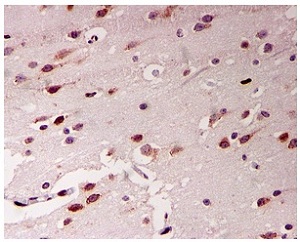
ab126745, at 1/50 dilution, staining ADAR1 in paraffin-embedded Human brain tissue by Immunohistochemistry.
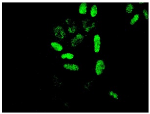
ab126745, at 1/50 dilution, staining ADAR1 in Hela cells by Immunofluorescence.
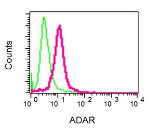
Flow cytometric analysis of permeabilized Ramos cells, staining ADAR1 (red) with ab126745. 1x10⁶ cells were collected and washed with blocking buffer. Cells were fixed with 2% paraformaldehyde, permeabilized with 1X FACS permeabilizing solution and blocked with blocking buffer for 30 minutes at room temperature. Cells were incubated with primary antibody (1/10) for 30 minutes at room temperature before a fluorescently-conjugated secondary antibody or 30 min at room temperature. A rabbit IgG was used as a negative control (green).