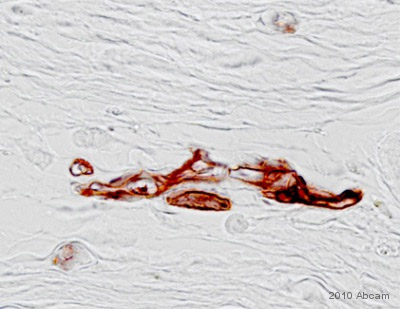
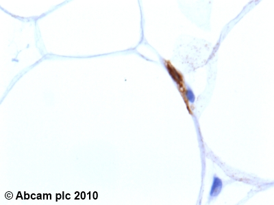
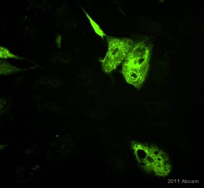
Anti-Adiponectin antibody [19F1]
| Name | Anti-Adiponectin antibody [19F1] |
|---|---|
| Supplier | Abcam |
| Catalog | ab22554 |
| Prices | $400.00 |
| Sizes | 100 µg |
| Host | Mouse |
| Clonality | Monoclonal |
| Isotype | IgG |
| Clone | 19F1 |
| Applications | ELISA ICC/IF ICC/IF ICC/IF IHC-P WB |
| Species Reactivities | Mouse, Rat, Rabbit, Human, Monkey |
| Antigen | Recombinant full length protein (Human) |
| Description | Mouse Monoclonal |
| Gene | ADIPOQ |
| Conjugate | Unconjugated |
| Supplier Page | Shop |
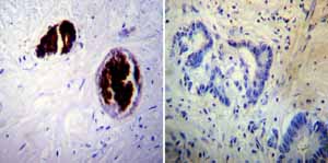
Product images
Product References
Identification of a distinct small cell population from human bone marrow reveals - Identification of a distinct small cell population from human bone marrow reveals
Wang J, Guo X, Lui M, Chu PJ, Yoo J, Chang M, Yen Y. PLoS One. 2014 Jan 17;9(1):e85112.
Adiponectin is sufficient, but not required, for exercise-induced increases in - Adiponectin is sufficient, but not required, for exercise-induced increases in
Ritchie IR, MacDonald TL, Wright DC, Dyck DJ. J Physiol. 2014 Jun 15;592(Pt 12):2653-65.
Adiponectin is not required for exercise training-induced improvements in glucose - Adiponectin is not required for exercise training-induced improvements in glucose
Ritchie IR, Wright DC, Dyck DJ. Physiol Rep. 2014 Sep 11;2(9). pii: e12146.
cAMP-responsive element binding protein: a vital link in embryonic hormonal - cAMP-responsive element binding protein: a vital link in embryonic hormonal
Schindler M, Fischer S, Thieme R, Fischer B, Santos AN. Endocrinology. 2013 Jun;154(6):2208-21.
Shotgun proteomics identifies serum fibronectin as a candidate diagnostic - Shotgun proteomics identifies serum fibronectin as a candidate diagnostic
Brown JK, Lauer KB, Ironmonger EL, Inglis NF, Bourne TH, Critchley HO, Horne AW. PLoS One. 2013 Jun 24;8(6):e66974.
Kruppel-like factor KLF9 regulates PPARgamma transactivation at the middle stage - Kruppel-like factor KLF9 regulates PPARgamma transactivation at the middle stage
Pei H, Yao Y, Yang Y, Liao K, Wu JR. Cell Death Differ. 2011 Feb;18(2):315-27.
Interleukin-1beta may mediate insulin resistance in liver-derived cells in - Interleukin-1beta may mediate insulin resistance in liver-derived cells in
Nov O, Kohl A, Lewis EC, Bashan N, Dvir I, Ben-Shlomo S, Fishman S, Wueest S, Konrad D, Rudich A. Endocrinology. 2010 Sep;151(9):4247-56.
Docosahexaenoic acid increases cellular adiponectin mRNA and secreted adiponectin - Docosahexaenoic acid increases cellular adiponectin mRNA and secreted adiponectin
Oster RT, Tishinsky JM, Yuan Z, Robinson LE. Appl Physiol Nutr Metab. 2010 Dec;35(6):783-9.
Adiponectin resistance precedes the accumulation of skeletal muscle lipids and - Adiponectin resistance precedes the accumulation of skeletal muscle lipids and
Mullen KL, Pritchard J, Ritchie I, Snook LA, Chabowski A, Bonen A, Wright D, Dyck DJ. Am J Physiol Regul Integr Comp Physiol. 2009 Feb;296(2):R243-51. doi:
Human CD34/CD90 ASCs are capable of growing as sphere clusters, producing high - Human CD34/CD90 ASCs are capable of growing as sphere clusters, producing high
De Francesco F, Tirino V, Desiderio V, Ferraro G, D'Andrea F, Giuliano M, Libondi G, Pirozzi G, De Rosa A, Papaccio G. PLoS One. 2009 Aug 6;4(8):e6537.
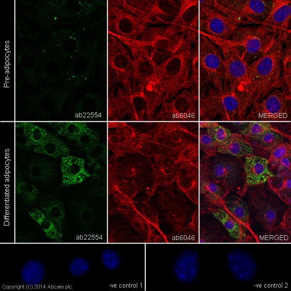
![Anti-Adiponectin antibody [19F1] (ab22554) at 1 µg/ml + 3T3-L1 (Mouse embryonic fibroblast - adipose like cell line) Nuclear Lysate (ab14632)](http://www.bioprodhub.com/system/product_images/ab_products/2/sub_1/3194_ab22554.jpg)
