![All lanes : Anti-AKT1/2/3 antibody [EPR17671] (ab185633) at 1/5000 dilutionLane 1 : AKT3 recombinant protein fragment (His-Tag®): aa351-479Lane 2 : AKT2 recombinant protein fragment (His-Tag®): aa282-481Lane 3 : AKT1 recombinant protein fragment (His-Tag®): aa281-480Lysates/proteins at 0.01 µg per lane.SecondaryAnti-Rabbit IgG (HRP), specific to the non-reduced form of IgG at 1/1000 dilution](http://www.bioprodhub.com/system/product_images/ab_products/2/sub_1/4371_ab185633-237228-wb-1.jpg)
All lanes : Anti-AKT1/2/3 antibody [EPR17671] (ab185633) at 1/5000 dilutionLane 1 : AKT3 recombinant protein fragment (His-Tag®): aa351-479Lane 2 : AKT2 recombinant protein fragment (His-Tag®): aa282-481Lane 3 : AKT1 recombinant protein fragment (His-Tag®): aa281-480Lysates/proteins at 0.01 µg per lane.SecondaryAnti-Rabbit IgG (HRP), specific to the non-reduced form of IgG at 1/1000 dilution
![Anti-AKT1/2/3 antibody [EPR17671] (ab185633) at 1/2000 dilution + A549 (Human lung carcinoma) whole cell lysates at 20 µgSecondaryGoat Anti-Rabbit IgG, (H+L),Peroxidase conjugated at 1/1000 dilution](http://www.bioprodhub.com/system/product_images/ab_products/2/sub_1/4372_ab185633-237229-wb-2.jpg)
Anti-AKT1/2/3 antibody [EPR17671] (ab185633) at 1/2000 dilution + A549 (Human lung carcinoma) whole cell lysates at 20 µgSecondaryGoat Anti-Rabbit IgG, (H+L),Peroxidase conjugated at 1/1000 dilution
![All lanes : Anti-AKT1/2/3 antibody [EPR17671] (ab185633) at 1/2000 dilutionLane 1 : Human fetal brain lysatesLane 2 : Human fetal kidney lysatesLysates/proteins at 10 µg per lane.SecondaryAnti-Rabbit IgG (HRP), specific to the non-reduced form of IgG at 1/1000 dilution](http://www.bioprodhub.com/system/product_images/ab_products/2/sub_1/4373_ab185633-237230-wb-3.jpg)
All lanes : Anti-AKT1/2/3 antibody [EPR17671] (ab185633) at 1/2000 dilutionLane 1 : Human fetal brain lysatesLane 2 : Human fetal kidney lysatesLysates/proteins at 10 µg per lane.SecondaryAnti-Rabbit IgG (HRP), specific to the non-reduced form of IgG at 1/1000 dilution
![All lanes : Anti-AKT1/2/3 antibody [EPR17671] (ab185633) at 1/2000 dilutionLane 1 : Mouse brain lysatesLane 2 : Rat brain lysatesLane 3 : Rat heart lysatesLysates/proteins at 10 µg per lane.SecondaryAnti-Rabbit IgG (HRP), specific to the non-reduced form of IgG at 1/1000 dilution](http://www.bioprodhub.com/system/product_images/ab_products/2/sub_1/4374_ab185633-237231-wb-4.jpg)
All lanes : Anti-AKT1/2/3 antibody [EPR17671] (ab185633) at 1/2000 dilutionLane 1 : Mouse brain lysatesLane 2 : Rat brain lysatesLane 3 : Rat heart lysatesLysates/proteins at 10 µg per lane.SecondaryAnti-Rabbit IgG (HRP), specific to the non-reduced form of IgG at 1/1000 dilution
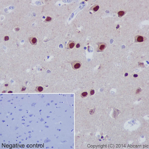
Immunohistochemical analysis of paraffin-embedded Human cerebral cortex tissue labeling AKT1/2/3 with ab185633 at 1/400 dilution, followed by Goat Anti-Rabbit IgG H&L (HRP) (ab97051) secondary antibody at 1/500 dilution. Nuclear and weak cytoplasmic staining on neurons of the Human cerebral cortex is observed. Counter stained with Hematoxylin.Negative control: Used PBS instead of primary antibody, secondary antibody is Goat Anti-Rabbit IgG H&L (HRP) (ab97051) at 1/500 dilution.
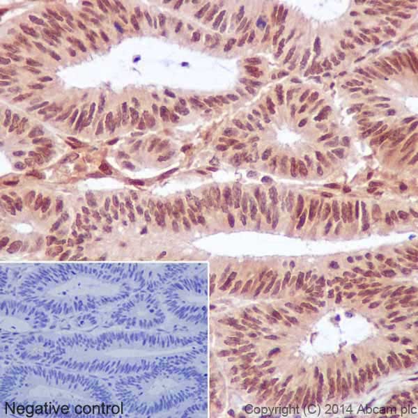
Immunohistochemical analysis of paraffin-embedded Human adenocarcinoma of colon tissue labeling AKT1/2/3 with ab185633 at 1/400 dilution, followed by Goat Anti-Rabbit IgG H&L (HRP) (ab97051) secondary antibody at 1/500 dilution. Nuclear and weak cytoplasmic staining on Human adenocarcinoma of colon is observed. Counter stained with Hematoxylin.Negative control: Used PBS instead of primary antibody, secondary antibody is Goat Anti-Rabbit IgG H&L (HRP) (ab97051) at 1/500 dilution.
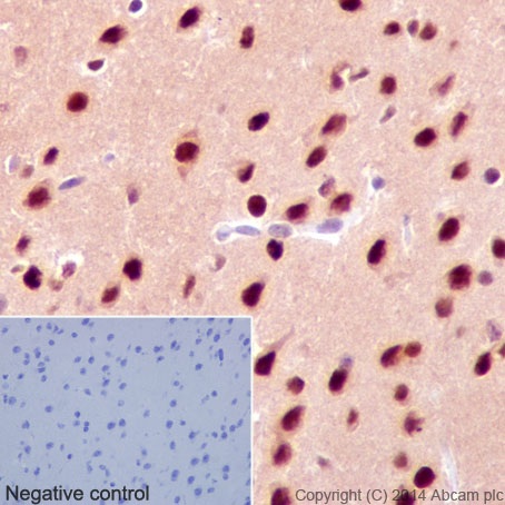
Immunohistochemical analysis of paraffin-embedded Mouse cerebral cortex tissue labeling AKT1/2/3 with ab185633 at 1/400 dilution, followed by Goat Anti-Rabbit IgG H&L (HRP) (ab97051) secondary antibody at 1/500 dilution. Nuclear and weak cytoplasmic staining on neurons of the mouse cerebral cortex is observed. Counter stained with Hematoxylin.Negative control: Used PBS instead of primary antibody, secondary antibody is Goat Anti-Rabbit IgG H&L (HRP) (ab97051) at 1/500 dilution.
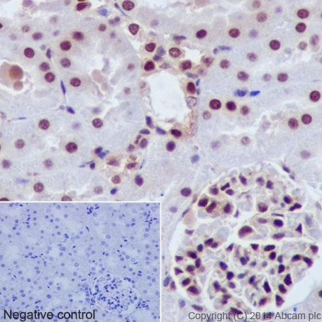
Immunohistochemical analysis of paraffin-embedded Rat kidney tissue labeling AKT1/2/3 with ab185633 at 1/400 dilution, followed by Goat Anti-Rabbit IgG H&L (HRP) (ab97051) secondary antibody at 1/500 dilution. Nuclear and weak cytoplasmic staining on rat kidney is observed. Counter stained with Hematoxylin.Negative control: Used PBS instead of primary antibody, secondary antibody is Goat Anti-Rabbit IgG H&L (HRP) (ab97051) at 1/500 dilution.
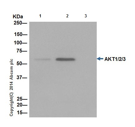
AKT1/2/3 was immunoprecipitated from 1mg of A549 (Human lung carcinoma) whole cell extract with ab185633 at 1/50 dilution. Western blot was performed from the immunoprecipitate using ab185633 at 1/1000 dilution. Anti-Rabbit IgG (HRP), specific to the non-reduced form of IgG, was used as secondary antibody at 1/1500 dilution.Lane 1: A549 whole cell extract 10 µg (Input). Lane 2: ab185633 IP in A549 whole cell extract. Lane 3: Rabbit monoclonal IgG (ab172730) instead of ab185633 in A549 whole cell extract.Blocking and dilution buffer and concentration: 5% NFDM/TBST.
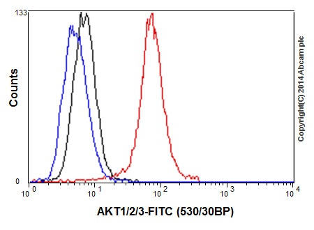
Flow cytometric analysis of 2% paraformaldehyde-fixed A549 (Human lung carcinoma) cells labeling AKT1/2/3 with ab185633 at 1/50 dilution (red) compared with a rabbit monoclonal IgG isotype control (black) and an unlabelled control (cells without incubation with primary antibody and secondary antibody; blue). Goat anti rabbit IgG (FITC) at 1/150 dilution was used as the secondary antibody.