Immunofluorescent analysis of AKT2 (green) showing staining in the cytoplasm and nucleus of C2C12 cells (right) compared to a negative control without primary antibody (left). Formalin-fixed cells were permeabilized with 0.1% Triton X-100 in TBS for 5-10 minutes and blocked with 3% BSA-PBS for 30 minutes at room temperature. Cells were probed with an AKT2 monoclonal antibody (ab175354) in 3% BSA-PBS at a dilution of 1:20 and incubated overnight at 4 ºC in a humidified chamber. Cells were washed with PBST and incubated with a DyLight-conjugated secondary antibody in PBS at room temperature in the dark. F-actin (red) was stained with a flourescent red phalloidin and nuclei (blue) were stained with Hoechst or DAPI. Images were taken at a magnification of 60x.

Immunofluorescent analysis of AKT2 (green) showing staining in the cytoplasm and nucleus of Hela cells (right) compared to a negative control without primary antibody (left). Formalin-fixed cells were permeabilized with 0.1% Triton X-100 in TBS for 5-10 minutes and blocked with 3% BSA-PBS for 30 minutes at room temperature. Cells were probed with an AKT2 monoclonal antibody (ab175354) in 3% BSA-PBS at a dilution of 1:20 and incubated overnight at 4 ºC in a humidified chamber. Cells were washed with PBST and incubated with a DyLight-conjugated secondary antibody in PBS at room temperature in the dark. F-actin (red) was stained with a flourescent red phalloidin and nuclei (blue) were stained with Hoechst or DAPI. Images were taken at a magnification of 60x.

Immunofluorescent analysis of AKT2 (green) showing staining in the cytoplasm and nucleus of MCF-7 cells (right) compared to a negative control without primary antibody (left). Formalin-fixed cells were permeabilized with 0.1% Triton X-100 in TBS for 5-10 minutes and blocked with 3% BSA-PBS for 30 minutes at room temperature. Cells were probed with an AKT2 monoclonal antibody (ab175354) in 3% BSA-PBS at a dilution of 1:20 and incubated overnight at 4 ºC in a humidified chamber. Cells were washed with PBST and incubated with a DyLight-conjugated secondary antibody in PBS at room temperature in the dark. F-actin (red) was stained with a flourescent red phalloidin and nuclei (blue) were stained with Hoechst or DAPI. Images were taken at a magnification of 60x.

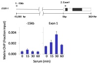
Chromatin immunoprecipitation analysis of Akt1 and Akt2 was performed using cross-linked chromatin from 1 x 10⁶ HCT116 colon carcinoma cells treated with serum for 0, 15, 30, and 60 minutes. Immunoprecipitation was performed with 1.0ul/100ul well volume of an Atk1 monoclonal antibody and an Akt2 monoclonal antibody (ab175354). Chromatin aliquots from ~1 x 10⁵ cells were used per ChIP pull-down. Quantitative PCR data were done in quadruplicate using 1ul of eluted DNA in 2ul SYBR real-time PCR reactions containing primers to amplify -15kb upstream of the Egr1 gene or exon-1 of Egr1. PCR calibration curves were generated for each primer pair from a dilution series of sheared total genomic DNA. Quantitation of immunoprecipitated chromatin is presented as signal relative to the total amount of input chromatin. Results represent the mean +/- SEM for three experiments. A schematic representation of the Egr-1 locus is shown above the data where boxes represent exons (black boxes = translated

Immunohistochemical analysis of deparaffinized Human Esophageal cancer tissue labeling AKT2 with ab175354 at 1/200 dilution. Detection was performed using a goat anti-mouse HRP secondary antibody followed by colorimetric detection using DAB substrate.
![All lanes : Anti-AKT2 antibody [4H7] - ChIP Grade (ab175354) at 1/1000 dilutionLane 1 : MCF7 whole cell lysateLane 2 : HeLa whole cell lysateLane 3 : HepG2 whole cell lysateLane 4 : A549 whole cell lysateLane 5 : 293T whole cell lysateLane 6 : Jurkat whole cell lysateLane 7 : A431 whole cell lysateLane 8 : U2OS whole cell lysateLane 9 : COS7 whole cell lysateLane 10 : 3T3 L1 whole cell lysateLane 11 : NRK whole cell lysateLysates/proteins at 25 µg per lane.Secondarygoat anti-mouse-HRP at 1/20000 dilutiondeveloped using the ECL technique](http://www.bioprodhub.com/system/product_images/ab_products/2/sub_1/4498_ab175354-204842-ab175354.jpg)
All lanes : Anti-AKT2 antibody [4H7] - ChIP Grade (ab175354) at 1/1000 dilutionLane 1 : MCF7 whole cell lysateLane 2 : HeLa whole cell lysateLane 3 : HepG2 whole cell lysateLane 4 : A549 whole cell lysateLane 5 : 293T whole cell lysateLane 6 : Jurkat whole cell lysateLane 7 : A431 whole cell lysateLane 8 : U2OS whole cell lysateLane 9 : COS7 whole cell lysateLane 10 : 3T3 L1 whole cell lysateLane 11 : NRK whole cell lysateLysates/proteins at 25 µg per lane.Secondarygoat anti-mouse-HRP at 1/20000 dilutiondeveloped using the ECL technique

Immunoprecipitation of AKT2 was performed on HeLa cells. The antigen:antibody complex was formed by incubating 750 µg whole cell lysate with 2 µg of ab175354. WB detection used ab175354 at 1/1000 dilution.

Immunohistochemical analysis of deparaffinized normal Human Medulla Oblongata tissue labeling AKT2 with ab175354 at 1/200 dilution. Detection was performed using a goat anti-mouse HRP secondary antibody followed by colorimetric detection using DAB substrate.
![All lanes : Anti-AKT2 antibody [4H7] - ChIP Grade (ab175354) at 1/1000 dilutionLane 1 : Non-transfected U2OS cellsLane 2 : AKT2 transfected U2OS cellsLysates/proteins at 25 per lane.Secondarygoat anti-mouse-HRP at 1/20000 dilution](http://www.bioprodhub.com/system/product_images/ab_products/2/sub_1/4501_ab175354-204843-ab175354b.jpg)
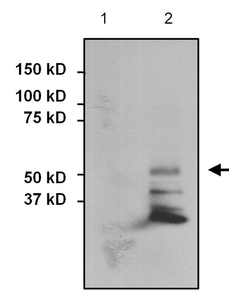
All lanes : Anti-AKT2 antibody [4H7] - ChIP Grade (ab175354) at 1/1000 dilutionLane 1 : Non-transfected U2OS cellsLane 2 : AKT2 transfected U2OS cellsLysates/proteins at 25 per lane.Secondarygoat anti-mouse-HRP at 1/20000 dilution


![All lanes : Anti-AKT2 antibody [4H7] - ChIP Grade (ab175354) at 1/1000 dilutionLane 1 : MCF7 whole cell lysateLane 2 : HeLa whole cell lysateLane 3 : HepG2 whole cell lysateLane 4 : A549 whole cell lysateLane 5 : 293T whole cell lysateLane 6 : Jurkat whole cell lysateLane 7 : A431 whole cell lysateLane 8 : U2OS whole cell lysateLane 9 : COS7 whole cell lysateLane 10 : 3T3 L1 whole cell lysateLane 11 : NRK whole cell lysateLysates/proteins at 25 µg per lane.Secondarygoat anti-mouse-HRP at 1/20000 dilutiondeveloped using the ECL technique](http://www.bioprodhub.com/system/product_images/ab_products/2/sub_1/4498_ab175354-204842-ab175354.jpg)


![All lanes : Anti-AKT2 antibody [4H7] - ChIP Grade (ab175354) at 1/1000 dilutionLane 1 : Non-transfected U2OS cellsLane 2 : AKT2 transfected U2OS cellsLysates/proteins at 25 per lane.Secondarygoat anti-mouse-HRP at 1/20000 dilution](http://www.bioprodhub.com/system/product_images/ab_products/2/sub_1/4501_ab175354-204843-ab175354b.jpg)