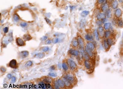
ab32816 (4 µg/ml) staining alpha actinin in human lung, using an automated system (DAKO Autostainer Plus). Using this protocol there is strong staining of both cytoplasm and nuclei of the bronchial epithelium and resident macrophage cells.Sections were rehydrated and antigen retrieved with the Dako 3 in 1 AR buffer EDTA pH 9.0 in a DAKO PT link. Slides were peroxidase blocked in 3% H2O2 in methanol for 10 mins. They were then blocked with Dako Protein block for 10 minutes (containing casein 0.25% in PBS) then incubated with primary antibody for 20 min and detected with Dako envision flex amplification kit for 30 minutes. Colorimetric detection was completed with Diaminobenzidine for 5 minutes. Slides were counterstained with Haematoxylin and coverslipped under DePeX. Please note that, for manual staining, optimization of primary antibody concentration and incubation time is recommended. Signal amplification may be required.
![Overlay histogram showing HeLa cells stained with ab32816 (red line). The cells were fixed with 80% methanol (5 min) and then permeabilized with 0.1% PBS-Tween for 20 min. The cells were then incubated in 1x PBS / 10% normal goat serum / 0.3M glycine to block non-specific protein-protein interactions followed by the antibody (ab32816, 1μg/1x106 cells) for 30 min at 22°C. The secondary antibody used was a goat anti-mouse DyLight® 488 (IgG; H+L) (ab96879) at 1/500 dilution for 30 min at 22°C. Isotype control antibody (black line) was a mix of mouse IgG1 [ICIGG1], (ab91353, 1μg/1x106 cells), IgG2a [ICIGG2A], (ab91361, 1μg/1x106 cells), IgG2b [PLPV219], (ab91366, 1μg/1x106 cells), IgG3 [MG3-35], (ab18394, 1μg/1x106 cells) used under the same conditions. Acquisition of >5,000 events was performed.](http://www.bioprodhub.com/system/product_images/ab_products/2/sub_1/5558_alpha-Actinin-4-Primary-antibodies-ab32816-3.jpg)
Overlay histogram showing HeLa cells stained with ab32816 (red line). The cells were fixed with 80% methanol (5 min) and then permeabilized with 0.1% PBS-Tween for 20 min. The cells were then incubated in 1x PBS / 10% normal goat serum / 0.3M glycine to block non-specific protein-protein interactions followed by the antibody (ab32816, 1μg/1x106 cells) for 30 min at 22°C. The secondary antibody used was a goat anti-mouse DyLight® 488 (IgG; H+L) (ab96879) at 1/500 dilution for 30 min at 22°C. Isotype control antibody (black line) was a mix of mouse IgG1 [ICIGG1], (ab91353, 1μg/1x106 cells), IgG2a [ICIGG2A], (ab91361, 1μg/1x106 cells), IgG2b [PLPV219], (ab91366, 1μg/1x106 cells), IgG3 [MG3-35], (ab18394, 1μg/1x106 cells) used under the same conditions. Acquisition of >5,000 events was performed.
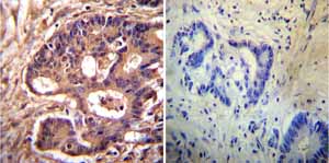
Immunohistochemistry was performed on both normal and cancer biopsies of deparaffinized Human colon carcinoma tissues. To expose target proteins heat induced antigen retrieval was performed using 10mM sodium citrate (pH6.0) buffer microwaved for 8-15 minutes. Following antigen retrieval tissues were blocked in 3% BSA-PBS for 30 minutes at room temperature. Tissues were then probed at a dilution of 1:200 with a mouse monoclonal antibody recognizing alpha Actinin 4 ab32816 or without primary antibody (negative control) overnight at 4°C in a humidified chamber. Tissues were washed extensively with PBST and endogenous peroxidase activity was quenched with a peroxidase suppressor. Detection was performed using a biotin-conjugated secondary antibody and SA-HRP followed by colorimetric detection using DAB. Tissues were counterstained with hematoxylin and prepped for mounting.
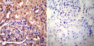
Immunohistochemistry was performed on both normal and cancer biopsies of deparaffinized Human kidney tissue tissues. To expose target proteins heat induced antigen retrieval was performed using 10mM sodium citrate (pH6.0) buffer microwaved for 8-15 minutes. Following antigen retrieval tissues were blocked in 3% BSA-PBS for 30 minutes at room temperature. Tissues were then probed at a dilution of 1:200 with a mouse monoclonal antibody recognizing alpha Actinin 4 ab32816 or without primary antibody (negative control) overnight at 4°C in a humidified chamber. Tissues were washed extensively with PBST and endogenous peroxidase activity was quenched with a peroxidase suppressor. Detection was performed using a biotin-conjugated secondary antibody and SA-HRP followed by colorimetric detection using DAB. Tissues were counterstained with hematoxylin and prepped for mounting.
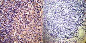
Immunohistochemistry was performed on both normal and cancer biopsies of deparaffinized Human tonsil tissue tissues. To expose target proteins heat induced antigen retrieval was performed using 10mM sodium citrate (pH6.0) buffer microwaved for 8-15 minutes. Following antigen retrieval tissues were blocked in 3% BSA-PBS for 30 minutes at room temperature. Tissues were then probed at a dilution of 1:200 with a mouse monoclonal antibody recognizing alpha Actinin 4 ab32816 or without primary antibody (negative control) overnight at 4°C in a humidified chamber. Tissues were washed extensively with PBST and endogenous peroxidase activity was quenched with a peroxidase suppressor. Detection was performed using a biotin-conjugated secondary antibody and SA-HRP followed by colorimetric detection using DAB. Tissues were counterstained with hematoxylin and prepped for mounting.
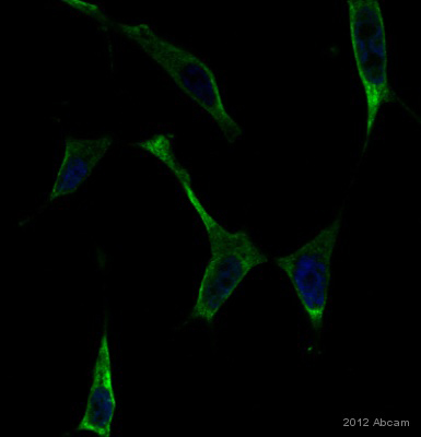
Immunofluorescence analysis of LNCaP cells, staining alpha Actinin 4 with ab32816. Cells were fixed with formaldehyde and blocked with 1% serum for 1 hour at 22°C. Samples were incubated with primary antibody (1/100 in 1% donkey serum in PBST) for 1 hour at 22°C. An undiluted DyLight®488-conjugated donkey anti-mouse polyclonal IgG was used as the secondary antibody. See Abreview

![Overlay histogram showing HeLa cells stained with ab32816 (red line). The cells were fixed with 80% methanol (5 min) and then permeabilized with 0.1% PBS-Tween for 20 min. The cells were then incubated in 1x PBS / 10% normal goat serum / 0.3M glycine to block non-specific protein-protein interactions followed by the antibody (ab32816, 1μg/1x106 cells) for 30 min at 22°C. The secondary antibody used was a goat anti-mouse DyLight® 488 (IgG; H+L) (ab96879) at 1/500 dilution for 30 min at 22°C. Isotype control antibody (black line) was a mix of mouse IgG1 [ICIGG1], (ab91353, 1μg/1x106 cells), IgG2a [ICIGG2A], (ab91361, 1μg/1x106 cells), IgG2b [PLPV219], (ab91366, 1μg/1x106 cells), IgG3 [MG3-35], (ab18394, 1μg/1x106 cells) used under the same conditions. Acquisition of >5,000 events was performed.](http://www.bioprodhub.com/system/product_images/ab_products/2/sub_1/5558_alpha-Actinin-4-Primary-antibodies-ab32816-3.jpg)


