Anti-alpha smooth muscle Actin antibody
| Name | Anti-alpha smooth muscle Actin antibody |
|---|---|
| Supplier | Abcam |
| Catalog | ab5694 |
| Prices | $401.00 |
| Sizes | 100 µg |
| Host | Rabbit |
| Clonality | Polyclonal |
| Isotype | IgG |
| Applications | IHC-F ICC/IF ICC/IF ICC/IF WB ELISA IHC-P IHC-F |
| Species Reactivities | Mouse, Rat, Chicken, Guinea Pig, Bovine, Dog, Human, Pig |
| Antigen | Synthetic peptide corresponding to Human alpha smooth muscle Actin |
| Description | Rabbit Polyclonal |
| Gene | ACTA2 |
| Conjugate | Unconjugated |
| Supplier Page | Shop |
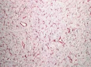
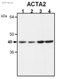
Product images
Product References
Telocytes are reduced during fibrotic remodelling of the colonic wall in - Telocytes are reduced during fibrotic remodelling of the colonic wall in
Manetti M, Rosa I, Messerini L, Ibba-Manneschi L. J Cell Mol Med. 2015 Jan;19(1):62-73.
Generation of the epicardial lineage from human pluripotent stem cells. - Generation of the epicardial lineage from human pluripotent stem cells.
Witty AD, Mihic A, Tam RY, Fisher SA, Mikryukov A, Shoichet MS, Li RK, Kattman SJ, Keller G. Nat Biotechnol. 2014 Oct;32(10):1026-35.
Interferon regulatory factor 8 modulates phenotypic switching of smooth muscle - Interferon regulatory factor 8 modulates phenotypic switching of smooth muscle
Zhang SM, Gao L, Zhang XF, Zhang R, Zhu LH, Wang PX, Tian S, Yang D, Chen K, Huang L, Zhang XD, Li H. Mol Cell Biol. 2014 Feb;34(3):400-14.
Cardiac-restricted overexpression or deletion of tissue inhibitor of matrix - Cardiac-restricted overexpression or deletion of tissue inhibitor of matrix
Yarbrough WM, Baicu C, Mukherjee R, Van Laer A, Rivers WT, McKinney RA, Prescott CB, Stroud RE, Freels PD, Zellars KN, Zile MR, Spinale FG. Am J Physiol Heart Circ Physiol. 2014 Sep 1;307(5):H752-61. doi:
Influenza A infection enhances antigen-induced airway inflammation and - Influenza A infection enhances antigen-induced airway inflammation and
Birmingham JM, Gillespie VL, Srivastava K, Li XM, Busse PJ. Clin Exp Allergy. 2014 Sep;44(9):1188-99.
Urinary semaphorin 3A correlates with diabetic proteinuria and mediates diabetic - Urinary semaphorin 3A correlates with diabetic proteinuria and mediates diabetic
Mohamed R, Ranganathan P, Jayakumar C, Nauta FL, Gansevoort RT, Weintraub NL, Brands M, Ramesh G. J Mol Med (Berl). 2014 Dec;92(12):1245-56.
Tissue injury and hypoxia promote malignant progression of prostate cancer by - Tissue injury and hypoxia promote malignant progression of prostate cancer by
Ammirante M, Shalapour S, Kang Y, Jamieson CA, Karin M. Proc Natl Acad Sci U S A. 2014 Oct 14;111(41):14776-81. doi:
Pattern formation of an epithelial tubule by mechanical instability during - Pattern formation of an epithelial tubule by mechanical instability during
Hirashima T. Cell Rep. 2014 Nov 6;9(3):866-73.
VEGF receptors mediate hypoxic remodeling of adult ovine carotid arteries. - VEGF receptors mediate hypoxic remodeling of adult ovine carotid arteries.
Adeoye OO, Bouthors V, Hubbell MC, Williams JM, Pearce WJ. J Appl Physiol (1985). 2014 Oct 1;117(7):777-87. doi:
PDGF receptor-alpha promotes TGF-beta signaling in hepatic stellate cells via - PDGF receptor-alpha promotes TGF-beta signaling in hepatic stellate cells via
Liu C, Li J, Xiang X, Guo L, Tu K, Liu Q, Shah VH, Kang N. Am J Physiol Gastrointest Liver Physiol. 2014 Oct 1;307(7):G749-59. doi:


