![Standard Curve for Apolipoprotein A I (Analyte: ab50239) dilution range 1pg/ml to 1ug/ml using Capture Antibody Mouse monoclonal [1409] to Apolipoprotein A I (ab20918) at 1ug/ml and Detector Antibody Rabbit polyclonal to Apolipoprotein A I (ab64308) at 0.5ug/ml](http://www.bioprodhub.com/system/product_images/ab_products/2/sub_1/8395_Apolipoprotein-A-I-Primary-antibodies-ab64308-8.jpg)
Standard Curve for Apolipoprotein A I (Analyte: ab50239) dilution range 1pg/ml to 1ug/ml using Capture Antibody Mouse monoclonal [1409] to Apolipoprotein A I (ab20918) at 1ug/ml and Detector Antibody Rabbit polyclonal to Apolipoprotein A I (ab64308) at 0.5ug/ml

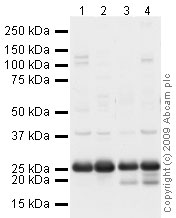
All lanes : Anti-Apolipoprotein A I antibody (ab64308) at 1 µg/mlLane 1 : Testis (Human) Tissue Lysate - adult normal tissue (ab30257)Lane 2 : Ovary (Human) Tissue Lysate - adult normal tissue (ab30222)Lane 3 : Lung (Human) Tissue Lysate - adult normal tissueLane 4 : Thymus (Human) Tissue Lysate - adult normal tissue (ab30146)Lysates/proteins at 10 µg per lane.SecondaryGoat polyclonal to Rabbit IgG - H&L - Pre-Adsorbed (HRP) at 1/3000 dilutionPerformed under reducing conditions.

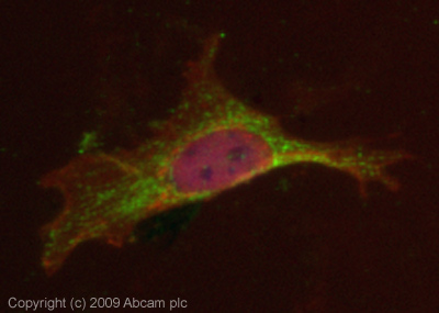
ICC/IF image of ab64308 stained HeLa cells. The cells were 4% PFA fixed (10 min) and then incubated in 1%BSA / 10% normal Goat serum / 0.3M glycine in 0.1% PBS-Tween for 1h to permeabilise the cells and block non-specific protein-protein interactions. The cells were then incubated with the antibody (ab64308, 5µg/ml) overnight at +4°C. The secondary antibody (green) was Alexa Fluor® 488 Goat anti-Rabbit IgG (H+L) used at a 1/1000 dilution for 1h. Alexa Fluor® 594 WGA was used to label plasma membranes (red) at a 1/200 dilution for 1h. DAPI was used to stain the cell nuclei (blue). This antibody also gave a positive result in 4% PFA fixed (10 min) Hek293, HepG2, and MCF-7 cells at 5µg/ml.

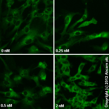
ab64308 staining ApoA1 in HepG1 cells treated with nicotinic acid (ab120145), by ICC/IF. Decrease in ApoA1 expression correlates with increased concentration of nicotinic acid, as described in literature.The cells were incubated at 37øC for 72h in media containing different concentrations of ab120145 ( nocotinic acid) in DMSO, fixed with 4% formaldehyde for 10 minutes at room temperature and blocked with PBS containing 10% goat serum, 0.3 M glycine, 1% BSA and 0.1% tween for 2h at room temperature. Staining of the treated cells with ab64308 (5 æg/ml) was performed overnight at 4øC in PBS containing 1% BSA and 0.1% tween. A DyLight 488 goat anti-rabbit polyclonal antibody (ab96899) at 1/250 dilution was used as the secondary antibody.
![Standard Curve for Apolipoprotein A I (Analyte: ab50239) dilution range 1pg/ml to 1ug/ml using Capture Antibody Mouse monoclonal [1409] to Apolipoprotein A I (ab20918) at 1ug/ml and Detector Antibody Rabbit polyclonal to Apolipoprotein A I (ab64308) at 0.5ug/ml](http://www.bioprodhub.com/system/product_images/ab_products/2/sub_1/8395_Apolipoprotein-A-I-Primary-antibodies-ab64308-8.jpg)


