Anti-Apolipoprotein CIII antibody
| Name | Anti-Apolipoprotein CIII antibody |
|---|---|
| Supplier | Abcam |
| Catalog | ab21032 |
| Prices | $390.00 |
| Sizes | 100 µg |
| Host | Rabbit |
| Clonality | Polyclonal |
| Isotype | IgG |
| Applications | ELISA ICC/IF ICC/IF ELISA WB |
| Species Reactivities | Human, Monkey |
| Antigen | Full length protein (Human) |
| Description | Rabbit Polyclonal |
| Gene | APOC3 |
| Conjugate | Unconjugated |
| Supplier Page | Shop |
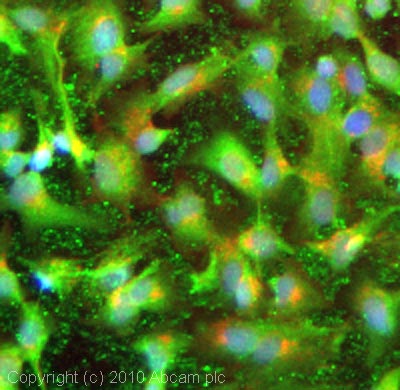
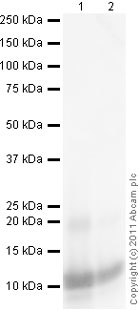
Product images
Product References
Probing isoform-specific functions of polypeptide GalNAc-transferases using zinc - Probing isoform-specific functions of polypeptide GalNAc-transferases using zinc
Schjoldager KT, Vakhrushev SY, Kong Y, Steentoft C, Nudelman AS, Pedersen NB, Wandall HH, Mandel U, Bennett EP, Levery SB, Clausen H. Proc Natl Acad Sci U S A. 2012 Jun 19;109(25):9893-8. doi:
Identification and quantification of differentially expressed proteins in plasma - Identification and quantification of differentially expressed proteins in plasma
Swarup V, Srivastava AK, Rajeswari MR. Neurosci Res. 2012 Jun;73(2):161-7.
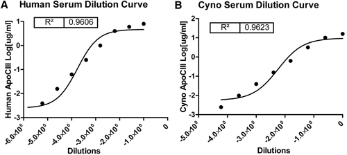
Development of a sensitive ELISA to quantify apolipoprotein CIII in nonhuman - Development of a sensitive ELISA to quantify apolipoprotein CIII in nonhuman
Wang Y, Song Z, Wagner JD, Pachuk C, Subramanian RR. J Lipid Res. 2011 Jun;52(6):1265-71.


