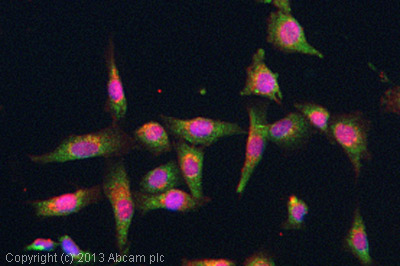
ICC/IF image of ab81355 stained SKNSH cells. The cells were 4% formaldehyde fixed (10 min) and then incubated in 1%BSA / 10% normal goat serum / 0.3M glycine in 0.1% PBS-Tween for 1h to permeabilise the cells and block non-specific protein-protein interactions. The cells were then incubated with the antibody (ab81355, 5µg/ml) overnight at +4°C. The secondary antibody (green) was ab96899, DyLight® 488 goat anti-rabbit IgG (H+L) used at a 1/250 dilution for 1h. Alexa Fluor® 594 WGA was used to label plasma membranes (red) at a 1/200 dilution for 1h. DAPI was used to stain the cell nuclei (blue) at a concentration of 1.43µM.

All lanes : Anti-Aquaporin 4 antibody (ab81355) at 1 µg/mlLane 1 : Tissue lysates prepared from Rat heart tissue.Lane 2 : Tissue lysates prepared from Rat kidney tissue.Lane 3 : Tissue lysates prepared from Rat brain tissue.Lane 4 : Tissue lysates prepared from Rat brain tissue.Lysates/proteins at 50 µg per lane.SecondaryHRP-conjugated Goat anti-rabbit IgG at 1/3000 dilution