Anti-Aryl hydrocarbon Receptor antibody (ab84833) at 1/200 dilution + HepG2 cell lysate

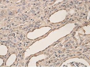
ab84833, at 1/200 dilution, staining Aryl hydrocarbon Receptor in formalin-fixed paraffin-embedded rat kidney by Immunohistochemistry.

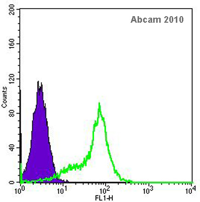
Flow cytometry using Aryl hydrocarbon Receptor antibody, ab84833. The histogram above shows ab84833 positive human embryonic stem cells (in green) and secondary only negative control (in purple).See Abreview

ab84833 staining Aryl hydrocarbon receptor in MDA-MB-231 cells treated with tranilast (ab120643), by ICC/IF. Increase in Aryl hydrocarbon receptor expression correlates with increased concentration of tranilast, as described in literature.The cells were incubated at 37°C for 24h in media containing different concentrations of ab120643 (telmisartan) in DMSO, fixed with 100% methanol for 5 minutes at -20°C and blocked with PBS containing 10% goat serum, 0.3 M glycine, 1% BSA and 0.1% tween for 2h at room temperature. Staining of the treated cells with ab84833 (5 µg/ml) was performed overnight at 4°C in PBS containing 1% BSA and 0.1% tween. A DyLight 488 goat anti-rabbit polyclonal antibody (ab96899) at 1/250 dilution was used as the secondary antibody. Nuclei were counterstained with DAPI and are shown in blue.

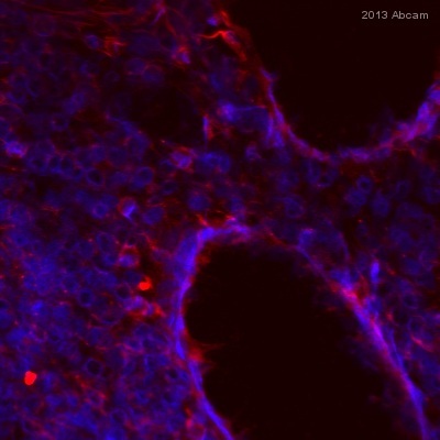
ab84833 staining Aryl hydrocarbon Receptor in Human colon tissue sections by Immunohistochemistry (IHC-Fr - frozen sections). Tissue was fixed with acetone and blocked with 10% sreum for 30 minutes at 21°C. Samples were incubated with primary antibody for 16 hours at 4°C. An undiluted Alexa fluor®555-conjugated Donkey anti-rabbit IgG polyclonal was used as the secondary antibody.See Abreview

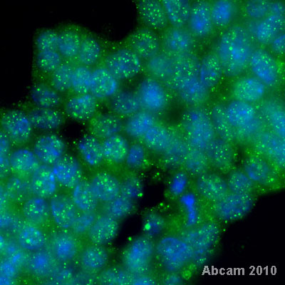
ab84833 staining Aryl hydrocarbon receptor protein on mouse embryonic stem cell by ICC/IF. The cells were paraformaldehyde fixed, permeabilized in 0.1% triton-x and blocked in 1% serum for 30 minutes at 21°C. The primary antibody was diluted 1/100 and incubated with sample for 2 hours at 21°C. An Alexa Fluor® 488 conjugated goat anti-rabbit IgG, diluted 1/100 was used as secondary.See Abreview


