Anti-Bax antibody
| Name | Anti-Bax antibody |
|---|---|
| Supplier | Abcam |
| Catalog | ab7977 |
| Prices | $400.00 |
| Sizes | 1 ml |
| Host | Rabbit |
| Clonality | Polyclonal |
| Isotype | IgG |
| Applications | IP IHC-P WB ICC/IF ICC/IF IHC-F |
| Species Reactivities | Mouse, Rat, Human, Hamster, Bovine |
| Antigen | Synthetic peptide corresponding to Human Bax aa 11-30 (N terminal) |
| Blocking Peptide | Human Bax peptide |
| Description | Rabbit Polyclonal |
| Gene | BAX |
| Conjugate | Unconjugated |
| Supplier Page | Shop |
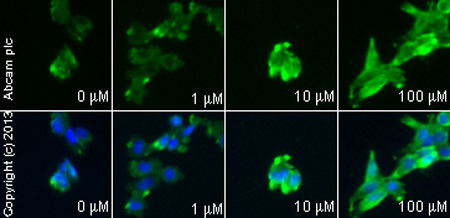
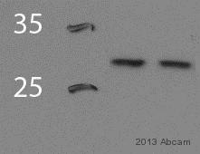
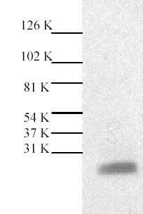
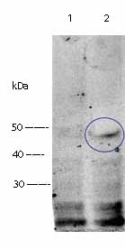
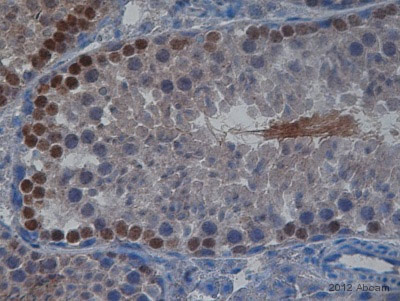
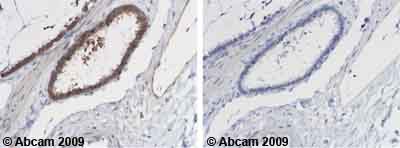
Product images
Product References
Preeclampsia is associated with alterations in the p53-pathway in villous - Preeclampsia is associated with alterations in the p53-pathway in villous
Sharp AN, Heazell AE, Baczyk D, Dunk CE, Lacey HA, Jones CJ, Perkins JE, Kingdom JC, Baker PN, Crocker IP. PLoS One. 2014 Jan 30;9(1):e87621.
Perivascular delivery of Notch 1 siRNA inhibits injury-induced arterial - Perivascular delivery of Notch 1 siRNA inhibits injury-induced arterial
Redmond EM, Liu W, Hamm K, Hatch E, Cahill PA, Morrow D. PLoS One. 2014 Jan 8;9(1):e84122.
Lactaptin induces p53-independent cell death associated with features of - Lactaptin induces p53-independent cell death associated with features of
Koval OA, Tkachenko AV, Fomin AS, Semenov DV, Nushtaeva AA, Kuligina EV, Zavjalov EL, Richter VA. PLoS One. 2014 Apr 7;9(4):e93921.
JWA regulates human esophageal squamous cell carcinoma and human esophageal cells - JWA regulates human esophageal squamous cell carcinoma and human esophageal cells
Lin J, Ma T, Jiang X, Ge Z, Ding W, Wu Y, Jiang G, Feng J, Cui G, Tan Y. Exp Ther Med. 2014 Jun;7(6):1767-1771. Epub 2014 Mar 28.
A novel androstenedione derivative induces ROS-mediated autophagy and attenuates - A novel androstenedione derivative induces ROS-mediated autophagy and attenuates
Liu Y, Zhao L, Ju Y, Li W, Zhang M, Jiao Y, Zhang J, Wang S, Wang Y, Zhao M, Zhang B, Zhao Y. Cell Death Dis. 2014 Aug 7;5:e1361.
Effect of phosphodiesterase-5 inhibition on apoptosis and beta amyloid load in - Effect of phosphodiesterase-5 inhibition on apoptosis and beta amyloid load in
Puzzo D, Loreto C, Giunta S, Musumeci G, Frasca G, Podda MV, Arancio O, Palmeri A. Neurobiol Aging. 2014 Mar;35(3):520-31. doi:
Gonadotropin-releasing hormone-regulated prohibitin mediates apoptosis of the - Gonadotropin-releasing hormone-regulated prohibitin mediates apoptosis of the
Savulescu D, Feng J, Ping YS, Mai O, Boehm U, He B, O'Malley BW, Melamed P. Mol Endocrinol. 2013 Nov;27(11):1856-70.
Antibody to mCLCA3 suppresses symptoms in a mouse model of asthma. - Antibody to mCLCA3 suppresses symptoms in a mouse model of asthma.
Song L, Liu D, Wu C, Wu S, Yang J, Ren F, Li Y. PLoS One. 2013 Dec 9;8(12):e82367.
The study of the Oxytropis kansuensis-induced apoptotic pathway in the cerebrum - The study of the Oxytropis kansuensis-induced apoptotic pathway in the cerebrum
Lu H, Zhang L, Wang SS, Wang WL, Zhao BY. BMC Vet Res. 2013 Oct 22;9:217.
Glycyrrhizin protects rat heart against ischemia-reperfusion injury through - Glycyrrhizin protects rat heart against ischemia-reperfusion injury through
Zhai CL, Zhang MQ, Zhang Y, Xu HX, Wang JM, An GP, Wang YY, Li L. Acta Pharmacol Sin. 2012 Dec;33(12):1477-87.
