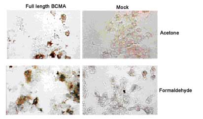
Immunostaining of HEK 293 cells transfected with a human BCMA expression plasmid (left panel), or mock transfected (right panel).Method: 3 days after transfection of cells with the indicated constructs, cells were fixed with acetone or 4% formaldehyde for 5min at RT. Slides were blocked with normal IgG, and incubated for 1 hour with 5µg/ml ab17323 in 1%BSA / 1x PBS. After washes in PBS, samples were incubated with the secondary antibody for 1 hour, washed in PBS and revealed with StreptABComplex/HRP (Vector) and AEC.

FACS analysis of cells with ab17323 to BCMA.Method: HEK 293 cells were mock transfected or transfected with an expression plasmid coding for human BCMA. Cells (5x10⁵) were incubated on ice for 30 min in 50µl FACS buffer (PBS, 5% fetal calf serum, 0.02% azide) containing 1µg/ml of ab17323 monoclonal antibody to human BCMA. After washing in FACS buffer, PE-conjugated antibody to rat IgG was added. Cells were incubated on ice for 30 min, washed and analyzed by flow cytometry.

Detection of endogenous human BCMA with ab17323 to BCMA (human)(Vicky-1) (second peak, to the right). Method: U266 cells (2x10⁵) were incubated on ice for 30 minutes with 0.2 µg of ab17323 or an isotype control in 25µl FACS buffer (PBS, 5% Fetal calf serum, 0.02% azide). The primary antibody was revealed with anti- Rat IgG (R-PE) and then analyzed by Flow Cytometry.