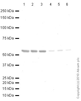
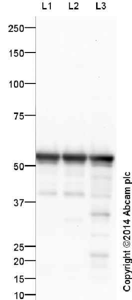
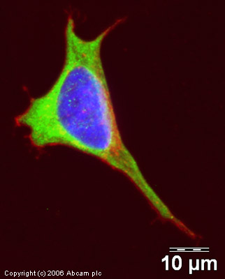
Anti-beta III Tubulin antibody
| Name | Anti-beta III Tubulin antibody |
|---|---|
| Supplier | Abcam |
| Catalog | ab18207 |
| Prices | $401.00 |
| Sizes | 100 µg |
| Host | Rabbit |
| Clonality | Polyclonal |
| Isotype | IgG |
| Applications | IHC-F FC IHC-P IHC-F ICC/IF ICC/IF WB |
| Species Reactivities | Mouse, Rat, Human, Pig, Marmoset |
| Antigen | Synthetic peptide conjugated to KLH derived from within residues 350 to the C-terminus of Human neuron specific beta III Tubulin |
| Blocking Peptide | Human beta III Tubulin peptide |
| Description | Rabbit Polyclonal |
| Gene | TUBB3 |
| Conjugate | Unconjugated |
| Supplier Page | Shop |
Product images
Product References
Complement inhibition and statins prevent fetal brain cortical abnormalities in a - Complement inhibition and statins prevent fetal brain cortical abnormalities in a
Pedroni SM, Gonzalez JM, Wade J, Jansen MA, Serio A, Marshall I, Lennen RJ, Girardi G. Biochim Biophys Acta. 2014 Jan;1842(1):107-15.
A disintegrin and metalloproteinases 10 and 17 modulate the immunogenicity of - A disintegrin and metalloproteinases 10 and 17 modulate the immunogenicity of
Wolpert F, Tritschler I, Steinle A, Weller M, Eisele G. Neuro Oncol. 2014 Mar;16(3):382-91.
Diffusion MR Microscopy of Cortical Development in the Mouse Embryo. - Diffusion MR Microscopy of Cortical Development in the Mouse Embryo.
Aggarwal M, Gobius I, Richards LJ, Mori S. Cereb Cortex. 2014 Feb 10.
A novel mouse model of Warburg Micro syndrome reveals roles for RAB18 in eye - A novel mouse model of Warburg Micro syndrome reveals roles for RAB18 in eye
Carpanini SM, McKie L, Thomson D, Wright AK, Gordon SL, Roche SL, Handley MT, Morrison H, Brownstein D, Wishart TM, Cousin MA, Gillingwater TH, Aligianis IA, Jackson IJ. Dis Model Mech. 2014 Jun;7(6):711-22.
Pleiotrophin antagonizes Brd2 during neuronal differentiation. - Pleiotrophin antagonizes Brd2 during neuronal differentiation.
Garcia-Gutierrez P, Juarez-Vicente F, Wolgemuth DJ, Garcia-Dominguez M. J Cell Sci. 2014 Jun 1;127(Pt 11):2554-64.
Morphine dependence in single enteric neurons from the mouse colon requires - Morphine dependence in single enteric neurons from the mouse colon requires
Smith TH, Ngwainmbi J, Hashimoto A, Dewey WL, Akbarali HI. Physiol Rep. 2014 Sep 4;2(9). pii: e12140.
Intrinsic innate immunity fails to control herpes simplex virus and vesicular - Intrinsic innate immunity fails to control herpes simplex virus and vesicular
Rosato PC, Leib DA. J Virol. 2014 Sep 1;88(17):9991-10001.
CFTR-deficient pigs display peripheral nervous system defects at birth. - CFTR-deficient pigs display peripheral nervous system defects at birth.
Reznikov LR, Dong Q, Chen JH, Moninger TO, Park JM, Zhang Y, Du J, Hildebrand MS, Smith RJ, Randak CO, Stoltz DA, Welsh MJ. Proc Natl Acad Sci U S A. 2013 Feb 19;110(8):3083-8. doi:
Focal Delivery of AAV2/1-transgenes Into the Rat Brain by Localized - Focal Delivery of AAV2/1-transgenes Into the Rat Brain by Localized
Alonso A, Reinz E, Leuchs B, Kleinschmidt J, Fatar M, Geers B, Lentacker I, Hennerici MG, de Smedt SC, Meairs S. Mol Ther Nucleic Acids. 2013 Feb 19;2:e73.
Lead induces similar gene expression changes in brains of gestationally exposed - Lead induces similar gene expression changes in brains of gestationally exposed
Sanchez-Martin FJ, Fan Y, Lindquist DM, Xia Y, Puga A. PLoS One. 2013 Nov 19;8(11):e80558.


