![beta III Tubulin was immunoprecipitated using 0.5mg Mouse Brain tissue, 5µg of Rabbit polyclonal to beta III Tubulin and 50µl of protein G magnetic beads (+). No antibody was added to the control (-).The antibody was incubated under agitation with Protein G beads for 10min, Mouse Brain tissue lysate diluted in RIPA buffer was added to each sample and incubated for a further 10min under agitation.Proteins were eluted by addition of 40µl SDS loading buffer and incubated for 10min at 70°C; 10µl of each sample was separated on a SDS PAGE gel, transferred to a nitrocellulose membrane, blocked with 5% BSA and probed with ab76287.Secondary: Mouse monoclonal [SB62a] Secondary Antibody to Rabbit IgG light chain (HRP) (ab99697).Band: 50kDa; beta III Tubulin,non specific - as present in control (lane 2); We are confident this was due to slight lane contamination and the band seen in the IP lane is our target of interest.](http://www.bioprodhub.com/system/product_images/ab_products/2/sub_1/14100_ab76287-198829-IPV023ab762872min.jpg)
beta III Tubulin was immunoprecipitated using 0.5mg Mouse Brain tissue, 5µg of Rabbit polyclonal to beta III Tubulin and 50µl of protein G magnetic beads (+). No antibody was added to the control (-).The antibody was incubated under agitation with Protein G beads for 10min, Mouse Brain tissue lysate diluted in RIPA buffer was added to each sample and incubated for a further 10min under agitation.Proteins were eluted by addition of 40µl SDS loading buffer and incubated for 10min at 70°C; 10µl of each sample was separated on a SDS PAGE gel, transferred to a nitrocellulose membrane, blocked with 5% BSA and probed with ab76287.Secondary: Mouse monoclonal [SB62a] Secondary Antibody to Rabbit IgG light chain (HRP) (ab99697).Band: 50kDa; beta III Tubulin,non specific - as present in control (lane 2); We are confident this was due to slight lane contamination and the band seen in the IP lane is our target of interest.

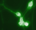
ab76287, at a 1/200 dilution, staining bea III Tubulin in chick dorsal root ganglion neurons by Immunoflurescence.
![beta III Tubulin was immunoprecipitated using 0.5mg Mouse Brain tissue, 5µg of Rabbit polyclonal to beta III Tubulin and 50µl of protein G magnetic beads (+). No antibody was added to the control (-).The antibody was incubated under agitation with Protein G beads for 10min, Mouse Brain tissue lysate diluted in RIPA buffer was added to each sample and incubated for a further 10min under agitation.Proteins were eluted by addition of 40µl SDS loading buffer and incubated for 10min at 70°C; 10µl of each sample was separated on a SDS PAGE gel, transferred to a nitrocellulose membrane, blocked with 5% BSA and probed with ab76287.Secondary: Mouse monoclonal [SB62a] Secondary Antibody to Rabbit IgG light chain (HRP) (ab99697).Band: 50kDa; beta III Tubulin,non specific - as present in control (lane 2); We are confident this was due to slight lane contamination and the band seen in the IP lane is our target of interest.](http://www.bioprodhub.com/system/product_images/ab_products/2/sub_1/14100_ab76287-198829-IPV023ab762872min.jpg)

