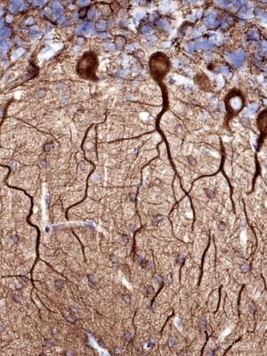
Immunohistochemistical detection of Neuron specific beta III Tubulin antibody using ab78078 on formaldehyde-fixed paraffin-embedded marmoset cerebellum sections. Antigen retrieval step: Heat mediated in Citric acid pH6 buffer. Permeabilization: No. Blocking step: 1% BSA for 10 mins @ rt°C. Primary Antibody used at 1/1000 incubated for 2 hours in TBS/BSA/azide. Secondary Antibody: anti mouse IgG conjugation to biotin (1/200).See Abreview
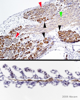
Immunohistochemical detection of Neuron specific beta III Tubulin using ab78078 on formaldehyde fixed dogfish/catshark PNS tissue sections. Antigen retrieval step: heat mediated in citric acid. Blocking: 1% BSA for 10 mins @ rt°C. Primary antibody ab78078 incubated at 1/1250 for 2 hours. Secondary Antibody: anti mouse IgG conjugated to biotin @ 1/200 This is a composite image of neuronal cell bodies and fibres; the upper image uses coloured arrowheads to indicate positive PNS components: cell bodies (black), L/S (green) and T/S (red) nerve fibres of what appear to be three Ganglia. Note that the L/S fibres do not appear as well-stained as the T/S fibres. The lower image shows what appears to be a linear sequence of single nerve cells and their processes within the core of a gill.
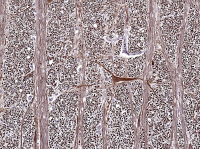
Immunohistochemistical detection of Neuron specific beta III Tubulin using antibody (ab78078) on formaldehyde-fixed paraffin-embedded human medulla oblongata sections. Antigen retrieval step: Heat mediated in citric acid HIER buffer. Permeabilization: No. Blocking step: 1% BSA for 10 mins @ rt°C. Primary antibody dilution 1/250 incubated for 2 hours in TBS/BSA/azide. Secondary antibody: anti Mouse IgGs conjugated to biotin (1/200). This section was cut from an anonymous autopsy P.wax block that is over 20 yrs old, so I would expect variable positivity when compared with more recently sampled tissues (giving higher dilution factors, such as I obtained with fresh mouse/rat CNS blocks.) Transverse-cut axons stain very intensely longitudinal nerve processes and cell bodies are not so heavily stained but are still easily seen.See Abreview
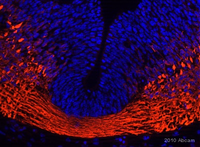
Immunohistochemistical detection of Neuron specific beta III Tubulin using antibody ab78078 on PFA fixed mouse embryo (image shows cervical spinal cord T/S). Primary Antibody Dilution: 1/1000 for 1 hour in TBS/BSA/azide. Secondary Antibody: anti Mouse IgG conjugated to Alexa Fluor® 594 (1/1000). The image submiteed here shows cervical spinal cord before closure.See Abreview
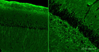
IHC-FoFr image of Beta III tubulin (ab78078) staining on rat hippocamus sections. The sections used came from animals perfused fixed with Paraformaldehyde 4%, in phosphate buffer 0.2M. Following postfixation in the same fixative overnight, the brains were cryoprotected in sucrose 30% overnight. Brains were then cut using a cryostat and the immunostainings were performed using the ‘free floating’ technique.See Abreview
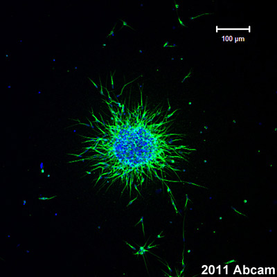
ab78078 staining a p8 mouse RMS neuroblast explant embedded in a 3D Matrigel matrix by ICC/IF. The explant was fixed with 4% paraformaldehyde for 45 minutes at room temperature and permeabilized with 0.3%Triton X. The explant was then blocked with 15% goat serum for 30 minutes at 21°C. The cultured explant was then stained with ab78078 at 1/2000 in 0.3% TritonX with 0.1x PBS and 15% goat serum for 16h at 4°C. An Alexa Fluro 488 goat anti-mouse polyclonal antibody at 1/1000 was used as the secondary antibody. Beta III tubulin (which stains the axons) can be observed in green and the nuclei can be obbserved in blue (DAPI).See Abreview
![All lanes : Anti-beta III Tubulin antibody [2G10] (ab78078) at 1 µg/mlLane 1 : Brain (Human) Tissue Lysate - adult normal tissue (ab29466)Lane 2 : Brain (Mouse) Tissue LysateLane 3 : Brain (Rat) Tissue LysateLysates/proteins at 10 µg per lane.SecondaryGoat Anti-Mouse IgG H&L (HRP) preadsorbed (ab97040) at 1/50000 dilutiondeveloped using the ECL techniquePerformed under reducing conditions.](http://www.bioprodhub.com/system/product_images/ab_products/2/sub_1/14118_ab78078-219498-WBAP16522621.jpg)
All lanes : Anti-beta III Tubulin antibody [2G10] (ab78078) at 1 µg/mlLane 1 : Brain (Human) Tissue Lysate - adult normal tissue (ab29466)Lane 2 : Brain (Mouse) Tissue LysateLane 3 : Brain (Rat) Tissue LysateLysates/proteins at 10 µg per lane.SecondaryGoat Anti-Mouse IgG H&L (HRP) preadsorbed (ab97040) at 1/50000 dilutiondeveloped using the ECL techniquePerformed under reducing conditions.
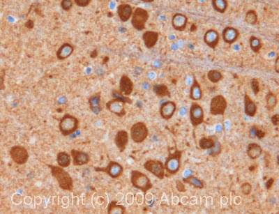
IHC image of ab78078 staining in mouse brain formalin fixed paraffin embedded tissue section, performed on a Leica BondTM system using the standard protocol F. The section was pre-treated using heat mediated antigen retrieval with sodium citrate buffer (pH6, epitope retrieval solution 1) for 20 mins. The section was then incubated with ab78078, 1µg/ml, for 15 mins at room temperature and detected using an HRP conjugated compact polymer system. DAB was used as the chromogen. The section was then counterstained with haematoxylin and mounted with DPX. For other IHC staining systems (automated and non-automated) customers should optimize variable parameters such as antigen retrieval conditions, primary antibody concentration and antibody incubation times.
![Immunohistochemistical detection of Neuron specific beta III Tubulin antibody [2G10] using ab78078 in formaldehyde-fixed paraffin-embedded quail embryo eye sections. Antigen retrieval step: heat mediated in citric acid pH6 buffer. Blocking step: 1% BSA for 10 mins @ rt°C Primary antibody incubated at 1/1250 for 2 hours in TBS/BSA/azide. Secondary antibody: anti-Mouse IgG conjugated to biotin used at 1/200. In this image, the lining cells of the hyaloid artery are positive. Excellent neural component specificity: even small fibres outside of the cartilage model of the orbit are clearly demonstrated.See Abreview](http://www.bioprodhub.com/system/product_images/ab_products/2/sub_1/14120_Neuron-specific-beta-III-Tubulin-Primary-antibodies-ab78078-16.jpg)
Immunohistochemistical detection of Neuron specific beta III Tubulin antibody [2G10] using ab78078 in formaldehyde-fixed paraffin-embedded quail embryo eye sections. Antigen retrieval step: heat mediated in citric acid pH6 buffer. Blocking step: 1% BSA for 10 mins @ rt°C Primary antibody incubated at 1/1250 for 2 hours in TBS/BSA/azide. Secondary antibody: anti-Mouse IgG conjugated to biotin used at 1/200. In this image, the lining cells of the hyaloid artery are positive. Excellent neural component specificity: even small fibres outside of the cartilage model of the orbit are clearly demonstrated.See Abreview
 used under the same conditions. Acquisition of >5,000 events was performed. This antibody gave a positive signal in SH-SY5Y cells fixed with 80% methanol/permeabilized in 0.1% PBS-Tween used under the same conditions.](http://www.bioprodhub.com/system/product_images/ab_products/2/sub_1/14121_Neuron-specific-beta-III-Tubulin-Primary-antibodies-ab78078-30.jpg)
Overlay histogram showing SH-SY5Y stained with ab78078 (red line). The cells were fixed with 4% paraformaldehyde (10 min) and then permeabilized with 0.1% PBS-Tween for 20 min. The cells were then incubated in 1x PBS / 10% normal goat serum / 0.3M glycine to block non-specific protein-protein interactions followed by the antibody (ab78078, 1µg/1x106 cells) for 30 min at 22°C. The secondary antibody used was DyLight® 488 goat anti-mouse IgG (H+L) (ab96879) at 1/500 dilution for 30 min at 22°C. Isotype control antibody (black line) was mouse IgG1 [ICIGG1](ab91353, 2µg/1x106 cells) used under the same conditions. Acquisition of >5,000 events was performed. This antibody gave a positive signal in SH-SY5Y cells fixed with 80% methanol/permeabilized in 0.1% PBS-Tween used under the same conditions.
![Neuron specific beta III Tubulin - Neuronal Marker was immunoprecipitated using 0.5mg Mouse Brain whole tissue lysate, 5µg of Mouse monoclonal [2G10] to Neuron specific beta III Tubulin - Neuronal Marker and 50µl of protein G magnetic beads (+). No antibody was added to the control (-). The antibody was incubated under agitation with Protein G beads for 10min, Mouse Brain whole tissue lysate lysate diluted in RIPA buffer was added to each sample and incubated for a further 10min under agitation.Proteins were eluted by addition of 40µl SDS loading buffer and incubated for 10min at 70oC; 10µl of each sample was separated on a SDS PAGE gel, transferred to a nitrocellulose membrane, blocked with 5% BSA and probed with ab78078.Secondary: Goat polyclonal to mouse IgG light chain specific (HRP) at 1/5000 dilution.Band: 50kDa: Neuron specific beta III Tubulin - Neuronal Marker.](http://www.bioprodhub.com/system/product_images/ab_products/2/sub_1/14122_Neuron-specific-beta-III-Tubulin-Primary-antibodies-ab78078-42.jpg)
Neuron specific beta III Tubulin - Neuronal Marker was immunoprecipitated using 0.5mg Mouse Brain whole tissue lysate, 5µg of Mouse monoclonal [2G10] to Neuron specific beta III Tubulin - Neuronal Marker and 50µl of protein G magnetic beads (+). No antibody was added to the control (-). The antibody was incubated under agitation with Protein G beads for 10min, Mouse Brain whole tissue lysate lysate diluted in RIPA buffer was added to each sample and incubated for a further 10min under agitation.Proteins were eluted by addition of 40µl SDS loading buffer and incubated for 10min at 70oC; 10µl of each sample was separated on a SDS PAGE gel, transferred to a nitrocellulose membrane, blocked with 5% BSA and probed with ab78078.Secondary: Goat polyclonal to mouse IgG light chain specific (HRP) at 1/5000 dilution.Band: 50kDa: Neuron specific beta III Tubulin - Neuronal Marker.
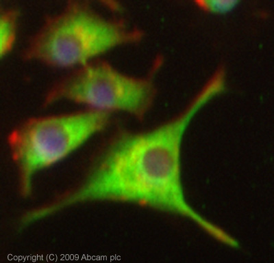
ICC/IF image of ab78078 stained PC12 cells. The cells were 4% PFA fixed (10 min) and then incubated in 1%BSA / 10% normal goat serum / 0.3M glycine in 0.1% PBS-Tween for 1h to permeabilise the cells and block non-specific protein-protein interactions. The cells were then incubated with the antibody (ab78078, 5µg/ml) overnight at +4°C. The secondary antibody (green) was Alexa Fluor® 488 goat anti-mouse IgG (H+L) used at a 1/1000 dilution for 1h. Alexa Fluor® 594 WGA was used to label plasma membranes (red) at a 1/200 dilution for 1h. DAPI was used to stain the cell nuclei (blue) at a concentration of 1.43µM. This antibody also gave a positive result in 100% methanol fixed (5 min) PC12 cells at 5µg/ml.
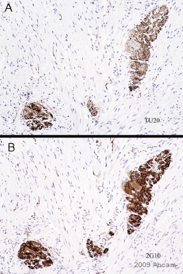
ab78078 staining Neuron specific beta III Tubulin in human colon tissue sections by Immunohistochemistry (Formalin/PFA-fixed paraffin-embedded sections). Tissue underwent formaldehyde fixation before heat mediated antigen retrieval in Citric acid pH6 and then blocking with 1% BSA was performed for 10 minutes at RT. The primary antibody was used at dilution at 1/200 and incubated with sample in TBS/BSA/azide for 2 hours. A Biotin conjugated goat polyclonal to mouse IgG was used at dilution at 1/200 as secondary antibody. Images A and B represent staining of the clone TU20 and 2G10 respectively in ganglia of Auerbach's plexus.See Abreview


![All lanes : Anti-beta III Tubulin antibody [2G10] (ab78078) at 1 µg/mlLane 1 : Brain (Human) Tissue Lysate - adult normal tissue (ab29466)Lane 2 : Brain (Mouse) Tissue LysateLane 3 : Brain (Rat) Tissue LysateLysates/proteins at 10 µg per lane.SecondaryGoat Anti-Mouse IgG H&L (HRP) preadsorbed (ab97040) at 1/50000 dilutiondeveloped using the ECL techniquePerformed under reducing conditions.](http://www.bioprodhub.com/system/product_images/ab_products/2/sub_1/14118_ab78078-219498-WBAP16522621.jpg)

![Immunohistochemistical detection of Neuron specific beta III Tubulin antibody [2G10] using ab78078 in formaldehyde-fixed paraffin-embedded quail embryo eye sections. Antigen retrieval step: heat mediated in citric acid pH6 buffer. Blocking step: 1% BSA for 10 mins @ rt°C Primary antibody incubated at 1/1250 for 2 hours in TBS/BSA/azide. Secondary antibody: anti-Mouse IgG conjugated to biotin used at 1/200. In this image, the lining cells of the hyaloid artery are positive. Excellent neural component specificity: even small fibres outside of the cartilage model of the orbit are clearly demonstrated.See Abreview](http://www.bioprodhub.com/system/product_images/ab_products/2/sub_1/14120_Neuron-specific-beta-III-Tubulin-Primary-antibodies-ab78078-16.jpg)
 used under the same conditions. Acquisition of >5,000 events was performed. This antibody gave a positive signal in SH-SY5Y cells fixed with 80% methanol/permeabilized in 0.1% PBS-Tween used under the same conditions.](http://www.bioprodhub.com/system/product_images/ab_products/2/sub_1/14121_Neuron-specific-beta-III-Tubulin-Primary-antibodies-ab78078-30.jpg)
![Neuron specific beta III Tubulin - Neuronal Marker was immunoprecipitated using 0.5mg Mouse Brain whole tissue lysate, 5µg of Mouse monoclonal [2G10] to Neuron specific beta III Tubulin - Neuronal Marker and 50µl of protein G magnetic beads (+). No antibody was added to the control (-). The antibody was incubated under agitation with Protein G beads for 10min, Mouse Brain whole tissue lysate lysate diluted in RIPA buffer was added to each sample and incubated for a further 10min under agitation.Proteins were eluted by addition of 40µl SDS loading buffer and incubated for 10min at 70oC; 10µl of each sample was separated on a SDS PAGE gel, transferred to a nitrocellulose membrane, blocked with 5% BSA and probed with ab78078.Secondary: Goat polyclonal to mouse IgG light chain specific (HRP) at 1/5000 dilution.Band: 50kDa: Neuron specific beta III Tubulin - Neuronal Marker.](http://www.bioprodhub.com/system/product_images/ab_products/2/sub_1/14122_Neuron-specific-beta-III-Tubulin-Primary-antibodies-ab78078-42.jpg)

