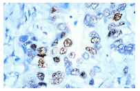
Rb (IF8): sc-102. Immunoperoxidase staining of formalin-fixed, paraffin-embedded human colon carcinoma tissue showing nuclear staining.
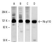
Western blot analysis of Rb p110 expression in A-431 whole cell lysate (A-C) and nuclear extract (D). Antibodies tested include Rb (C-15): sc-50 (A), Rb (C-15)-G: sc-50-G (B), Rb (IF8): sc-102 (C) and Rb (M-153): sc-7905 (D).
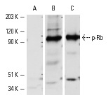
Western blot analysis of Rb phosphorylation in SK-LMS-1 whole cell lysate. Blots were probed with p-Rb (Ser 807/811)-R: sc-16670-R preincubated with cognate phosphorylated peptide (A) or unphosphorylated peptide (B) and Rb (IF8): sc-102 (C).
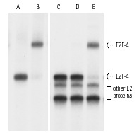
Gel supershift analysis of E2F proteins isolated by immunoprecipitation using either p107 (A,B) or Rb p110 (C-E) antibodies. E2F-4 released from p107/E2F complexes and incubated with
32P-labeled E2F binding sites specific oligonucleotide was supershifted by E2F-4 (C-108) X: sc-512 X antibody (B) as compared to control IgG (A). Similarly, E2F-4 released from Rb p110/E2F complexes was supershifted by E2F-4 (C-108) X: sc-512 X (E), but not by E2F-1 (KH95) X: sc-251 X (D) as compared to control IgG (C). Kindly provided by Masa-aki Ikeda, Ph.D., D.D.S.
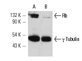
Rb siRNA (h): sc-29468. Western blot analysis of Rb expression in non-transfected control (A) and Rb siRNA transfected (B) HeLa cells. Blot probed with Rb (IF8): sc-102. γ Tubulin (C-11): sc-17787 used as specificity and loading control.
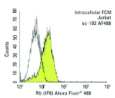
Rb (IF8) Alexa Fluor 488: sc-102 AF488. Intracellular FCM analysis of fixed and permeabilized Jurkat cells. Black line histogram represents the isotype control, normal mouse IgG
1: sc-3890.
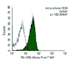
Rb (IF8) Alexa Fluor 647: sc-102 AF647. Intracellular FCM analysis of fixed and permeabilized Jurkat cells. Black line histogram represents the isotype control, normal mouse IgG
1: sc-24636.
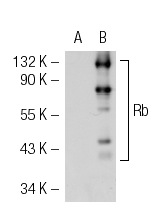
Rb (IF8): sc-102. Western blot analysis of Rb expression in non-transfected: sc-117752 (A) and human Rb transfected: sc-114014 (B) 293T whole cell lysates.
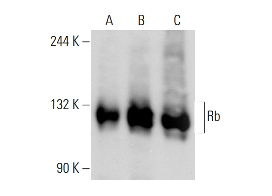
Rb (IF8): sc-102. Western blot analysis of Rb expression in non-transfected 293T: sc-117752 (A), human Rb transfected 293T: sc-159907 (B) and K-562 (C) whole cell lysates.
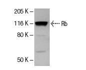
Rb (IF9): sc-102. Western blot analysis of Rb expression in K-562 whole cell lysate.
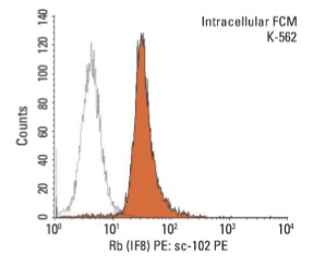
Rb (IF8) PE: sc-102 PE. Intracellular FCM analysis of fixed and permeabilized K-562 cells. Black line histogram represents the isotype control, normal mouse IgG
1: sc-2866.


