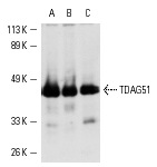
TDAG51 (RN-6E2): sc-23866. Western blot analysis of TDAG51 expression in Hep G2 (A), HUVEC (B) and HeLa (C) whole cell lysates.
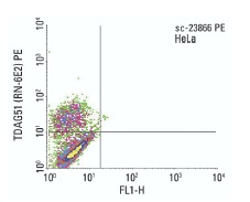
TDAG51 (RN-6E2) PE: sc-23866 PE. FCM analysis of HeLa cells. Quadrant markers were set based on the isotype control, normal mouse IgG
2a: sc-2867.
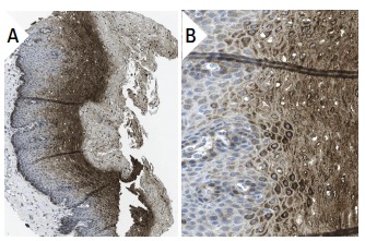
TDAG51 (RN-6E2): sc-23866. Immunoperoxidase staining of formalin fixed, paraffin-embedded human esophagus tissue showing cytoplasmic staining of squamous epithelial cells (low and high magnification). Kindly provided by The Swedish Human Protein Atlas (HPA) program.
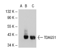
TDAG51 (RN-6E2): sc-23866. Western blot analysis of TDAG51 expression in non-transfected 293T: sc-117752 (A), human TDAG51 transfected 293T: sc-111445 (B) and Hep G2 (C) whole cell lysates.
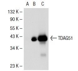
TDAG51 (RN-6E2): sc-23866. Western blot analysis of TDAG51 expression in non-transfected 293T: sc-117752 (A), mouse TDAG51 transfected 293T: sc-123964 (B) and Hep G2 (C) whole cell lysates.
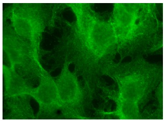
TDAG51 (RN-6E2): sc-23866. Immunofluorescence staining of methanol-fixed Hep G2 cells showing membrane localization.
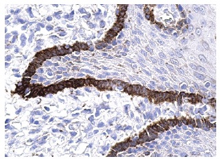
TDAG51 (RN-6E2): sc-23866. Immunoperoxidase staining of formalin fixed, paraffin-embedded human esophagus tissue showing cytoplasmic staining of basal squamous epithelial cells.


