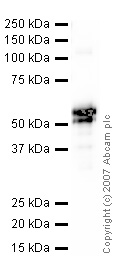
Anti-Carbonic Anhydrase IX antibody (ab10471) at 1 µg/ml + Carbonic Anhydrase IX recombinant protein at 0.1 µg/mlSecondaryGoat polyclonal to Rabbit IgG H&L (HRP) Pre-Adsorbed at 1/10000 dilutionPerformed under reducing conditions.
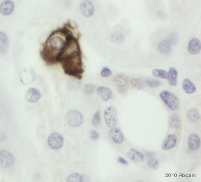
ab10471 staining Carbonic Anhydrase IX in Human kidney tissue sections by IHC-P (Formaldehyde-fixed, paraffin-embedded sections). Tissue was fixed with formaldehyde and blocked with 5 minutes of peroxidase block followed by 10 minutes of protein block at 20°C; antigen retrieval was heat mediated in retrieval solution. Samples were incubated with primary antibody (1/250 in antibody diluent) for 45 minutes at 20°C. An undiluted HRP-conjugated polymer goat anti-mouse/rabbit IgG polyclonal was used as secondary antibody.See Abreview

ICC/IF image of ab10471 stained DU145 cells. The cells were 100% methanol fixed (5 min) and then incubated in 1%BSA / 10% normal goat serum / 0.3M glycine in 0.1% PBS-Tween for 1h to permeabilise the cells and block non-specific protein-protein interactions. The cells were then incubated with the antibody ab10471 at 5µg/ml overnight at +4°C. The secondary antibody (green) was DyLight® 488 goat anti- rabbit (ab96899) IgG (H+L) used at a 1/1000 dilution for 1h. Alexa Fluor® 594 WGA was used to label plasma membranes (red) at a 1/200 dilution for 1h. DAPI was used to stain the cell nuclei (blue) at a concentration of 1.43µM.