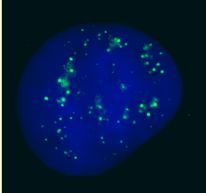
Immunofluorescent imaging of human cells (U2OS) with ab4189 reveals the expected discrete nuclear structure that is termed the PcG body corresponding to the known localisation of PC2. This corresponds to results seen in Satijn et al.IF was performed with a standard paraformaldehyde technique (fixed in PBS buffered PFH 4% for 5 minutes, permeabilised with 0.5% triton-PBS for 5 minutes, blocked with 5% milk / 0.2% tween for one hour. Primary antibody used at 1/200 in 5% milk / 0.2% TWEEN for one hour, secondary antibody for 30 minutes. All blocking and incubation steps carried out at 37 degrees C. Nuclei counterstained with Hoechst stain (blue).
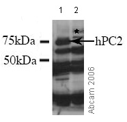 All lanes : Anti-CBX4 antibody (ab4189)Lane 1 : HEK 293 lysate over-expressing DDDDK tagged hPC2 Lane 2 : HEK 293 lysate