![All lanes : Anti-CC2D1A antibody [EPR18421] (ab191472) at 1/20000 dilutionLane 1 : Hela (Human epithelial cells from cervix adenocarcinoma) whole cell lysatesLane 2 : HEK-293 (Human epithelial cells from embryonic kidney) whole cell lysatesLane 3 : Jurkat (Human T cell leukemia cells from peripheral blood) whole cell lysatesLane 4 : MCF7 (Human breast adenocarcinoma cell line) whole cell lysatesLysates/proteins at 20 µg per lane.SecondaryGoat Anti-Rabbit IgG H&L (HRP) (ab97051) at 1/1000 dilution](http://www.bioprodhub.com/system/product_images/ab_products/2/sub_1/22167_ab191472-246121-wb-1.jpg)
All lanes : Anti-CC2D1A antibody [EPR18421] (ab191472) at 1/20000 dilutionLane 1 : Hela (Human epithelial cells from cervix adenocarcinoma) whole cell lysatesLane 2 : HEK-293 (Human epithelial cells from embryonic kidney) whole cell lysatesLane 3 : Jurkat (Human T cell leukemia cells from peripheral blood) whole cell lysatesLane 4 : MCF7 (Human breast adenocarcinoma cell line) whole cell lysatesLysates/proteins at 20 µg per lane.SecondaryGoat Anti-Rabbit IgG H&L (HRP) (ab97051) at 1/1000 dilution
![All lanes : Anti-CC2D1A antibody [EPR18421] (ab191472) at 1/5000 dilutionLane 1 : C6 (Rat glial tumor cells) whole cell lysateLane 2 : Raw264.7 (Mouse macrophage cells transformed with Abelson murine leukemia virus) whole cell lysateLane 3 : PC12 (Rat adrenal gland pheochromocytoma) whole cell lysateLane 4 : NIH 3T3 (Mouse embyro fibroblast cells) whole cell lysateLysates/proteins at 10 µg per lane.SecondaryGoat Anti-Rabbit IgG H&L (HRP) (ab97051) at 1/1000 dilution](http://www.bioprodhub.com/system/product_images/ab_products/2/sub_1/22168_ab191472-246120-wb-2.jpg)
All lanes : Anti-CC2D1A antibody [EPR18421] (ab191472) at 1/5000 dilutionLane 1 : C6 (Rat glial tumor cells) whole cell lysateLane 2 : Raw264.7 (Mouse macrophage cells transformed with Abelson murine leukemia virus) whole cell lysateLane 3 : PC12 (Rat adrenal gland pheochromocytoma) whole cell lysateLane 4 : NIH 3T3 (Mouse embyro fibroblast cells) whole cell lysateLysates/proteins at 10 µg per lane.SecondaryGoat Anti-Rabbit IgG H&L (HRP) (ab97051) at 1/1000 dilution
![Anti-CC2D1A antibody [EPR18421] (ab191472) at 1/20000 dilution + Mouse brain lysate at 10 µgSecondaryGoat Anti-Rabbit IgG H&L (HRP) (ab97051) at 1/1000 dilution](http://www.bioprodhub.com/system/product_images/ab_products/2/sub_1/22169_ab191472-246119-wb-3.jpg)
Anti-CC2D1A antibody [EPR18421] (ab191472) at 1/20000 dilution + Mouse brain lysate at 10 µgSecondaryGoat Anti-Rabbit IgG H&L (HRP) (ab97051) at 1/1000 dilution
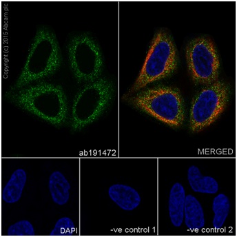
Immunofluorescent analysis of 4% paraformaldehyde-fixed, 0.1% Triton X-100 permeabilized HeLa (Human epithelial cells from cervix adenocarcinoma) cells labeling CC2D1A with ab191472 at 1/150 dilution, followed by Goat anti-rabbit IgG (Alexa Fluor® 488) (ab150077) secondary antibody at 1/1000 dilution (green).Confocal image showing cytoplasm staining on HeLa cell line.The nuclear counter stain is DAPI (blue).Tubulin is detected with ab7291 (anti-Tubulin mouse mAb) at 1/1000 dilution and ab150120 (AlexaFluor®594 Goat anti-Mouse secondary) at 1/1000 dilution (red).The negative controls are as follows:1. ab191472 at 1/150 dilution followed by ab150120 (AlexaFluor®594 Goat anti-Mouse secondary) at 1/1000 dilution.2. ab7291 (anti-Tubulin mouse mAb) at 1/1000 dilution followed by ab150077 (Alexa Fluor®488 Goat Anti-Rabbit IgG H&L) at 1/1000 dilution.
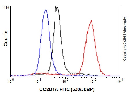
Flow cytometric analysis of 4% paraformaldehyde-fixed HeLa (Human epithelial cells from cervix adenocarcinoma) cells labeling CC2D1A with ab191472 at 1/250 dilution (red) compared with a rabbit monoclonal IgG isotype control (ab172730; black) and a unlabelled control (cells without incubation with primary antibody and secondary antibody; blue). Goat anti rabbit IgG (FITC) at 1/500 dilution was used as the secondary antibody.