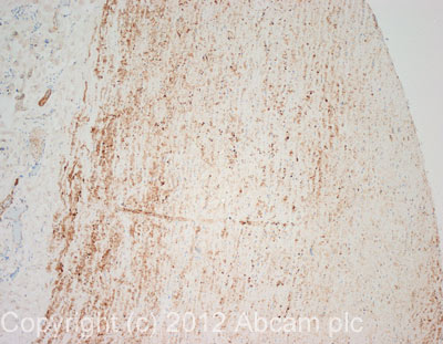
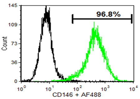
Anti-CD146 antibody [P1H12]
| Name | Anti-CD146 antibody [P1H12] |
|---|---|
| Supplier | Abcam |
| Catalog | ab24577 |
| Prices | $403.00 |
| Sizes | 100 µg |
| Host | Mouse |
| Clonality | Monoclonal |
| Isotype | IgG1 |
| Clone | P1H12 |
| Applications | ICC/IF ICC/IF IHC-P WB ELISA IHC-F IP FC |
| Species Reactivities | Human, Mouse, Dog |
| Antigen | Tissue/ cell preparation: human umbilical vein endothelial cells (HUVECs) |
| Description | Mouse Monoclonal |
| Gene | MCAM |
| Conjugate | Unconjugated |
| Supplier Page | Shop |
Product images
Product References
Transcript analysis reveals a specific HOX signature associated with positional - Transcript analysis reveals a specific HOX signature associated with positional
Toshner M, Dunmore BJ, McKinney EF, Southwood M, Caruso P, Upton PD, Waters JP, Ormiston ML, Skepper JN, Nash G, Rana AA, Morrell NW. PLoS One. 2014 Mar 20;9(3):e91334.
Isolation and characterization of mesenchymal progenitor cells from human orbital - Isolation and characterization of mesenchymal progenitor cells from human orbital
Chen SY, Mahabole M, Horesh E, Wester S, Goldberg JL, Tseng SC. Invest Ophthalmol Vis Sci. 2014 Jul 3;55(8):4842-52.
Perivascular-like cells contribute to the stability of the vascular network of - Perivascular-like cells contribute to the stability of the vascular network of
Mendes LF, Pirraco RP, Szymczyk W, Frias AM, Santos TC, Reis RL, Marques AP. PLoS One. 2012;7(7):e41051.
Activation of protease-activated receptor 2 induces VEGF independently of HIF-1. - Activation of protease-activated receptor 2 induces VEGF independently of HIF-1.
Rasmussen JG, Riis SE, Frobert O, Yang S, Kastrup J, Zachar V, Simonsen U, Fink T. PLoS One. 2012;7(9):e46087.
An isolated cryptic peptide influences osteogenesis and bone remodeling in an - An isolated cryptic peptide influences osteogenesis and bone remodeling in an
Agrawal V, Kelly J, Tottey S, Daly KA, Johnson SA, Siu BF, Reing J, Badylak SF. Tissue Eng Part A. 2011 Dec;17(23-24):3033-44.
Development of microfluidics as endothelial progenitor cell capture technology - Development of microfluidics as endothelial progenitor cell capture technology
Plouffe BD, Kniazeva T, Mayer JE Jr, Murthy SK, Sales VL. FASEB J. 2009 Oct;23(10):3309-14.
Circulating activated endothelial cells in sickle cell anemia. - Circulating activated endothelial cells in sickle cell anemia.
Solovey A, Lin Y, Browne P, Choong S, Wayner E, Hebbel RP. N Engl J Med. 1997 Nov 27;337(22):1584-90.

![All lanes : Anti-CD146 antibody [P1H12] (ab24577) at 5 µg/mlLane 1 : HUVEC (Human Umbilical Vein Endothelial Cell) Whole Cell Lysate Lane 2 : Blood Vessel: artery (Human) Membrane Lysate - adult normal tissue (ab28989)Lysates/proteins at 25 µg per lane.SecondaryGoat polyclonal Secondary Antibody to Mouse IgG - H&L (HRP), pre-adsorbed at 1/5000 dilutionPerformed under reducing conditions.](http://www.bioprodhub.com/system/product_images/ab_products/2/sub_1/23373_CD146-Primary-antibodies-ab24577-14.jpg)


