MCAM

Gene Symbol MCAM
Entrez Gene 4162
Alt Symbol CD146, MUC18
Species Human
Gene Type protein-coding
Description melanoma cell adhesion molecule
Other Description Gicerin|S-endo 1 endothelial-associated antigen|cell surface glycoprotein MUC18|cell surface glycoprotein P1H12|melanoma adhesion molecule|melanoma-associated antigen A32|melanoma-associated antigen MUC18
Swissprots Q6PHR3 Q59E86 Q6ZTR2 O95812 P43121
Accessions CAA48332 CCF76943 EAW67477 EAW67478 P43121 AB209925 BAD93162 AF089868 AAD17799 AJ297452 CAB97348 AK126303 BAC86520 AK128335 BAG54661 AK291571 BAF84260 AK312430 BAG35339 AU076908 BC056418 AAH56418 EU832794 ACE87805 GQ129330 ACT64464 M28882 AAA20922 M29277 AAA20824 NM_006500 NP_006491
Function Plays a role in cell adhesion, and in cohesion of the endothelial monolayer at intercellular junctions in vascular tissue. Its expression may allow melanoma cells to interact with cellular elements of the vascular system, thereby enhancing hematogeneous tumor spread. Could be an adhesion molecule active in neural crest cells during embryonic development. Acts as surface receptor that triggers tyrosine phosphorylation of FYN and PTK2/FAK1, and a transient increase in the intracellular calcium concentration. {ECO:0000269|PubMed:11036077, ECO:0000269|PubMed:8292890}.
Subcellular Location Membrane; Single-pass type I membrane protein.
Tissue Specificity Detected in endothelial cells in vascular tissue throughout the body. May appear at the surface of neural crest cells during their embryonic migration. Appears to be limited to vascular smooth muscle in normal adult tissues. Associated with tumor progression and the development of metastasis in human malignant melanoma. Expressed most strongly on metastatic lesions and advanced primary tumors and is only rarely detected in benign melanocytic nevi and thin primary melanomas with a low probability of metastasis.

MCAM (P1H12) Mouse mAb - 13475 from Cell Signaling Technology

Prices $246.00
Sizes 100 µl (10 western blots)
Host Mouse
Clonality Monoclonal
Clone P1H12
Western blot analysis of extracts from SK-MEL-28, HUVEC, and COLO 205 cells using MCAM (P1H12) Mouse mAb (upper) and GAPDH (D16H11) XP ® Rabbit mAb #5174 (lower). As expected, COLO 205 cells are negative for MCAM protein.

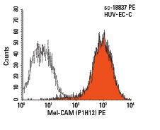
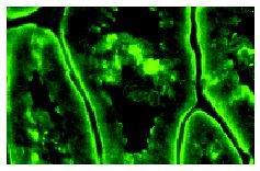
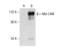
Mel-CAM (P1H12) - sc-18837 from Santa Cruz Biotechnology

Prices $279.00
Sizes 200 µg/ml
Host Mouse
Clonality Monoclonal
Mel-CAM (P1H12) PE: sc-18837 PE. FCM analysis of HUV-EC-C cells. Black line histogram represents the isotype control, normal mouse IgG<sub>1</sub>: sc-2866.

Mel-CAM (C-20) - sc-18942 from Santa Cruz Biotechnology

Prices $279.00
Sizes 200 µg/ml
Host Goat
Clonality Polyclonal
Mel-CAM (C-20): sc-18942. Western blot analysis of Mel-CAM expression in A-375 whole cell lysate.

Mel-CAM (H-62) - sc-28667 from Santa Cruz Biotechnology

Prices $279.00
Sizes 200 µg/ml
Host Rabbit
Clonality Polyclonal
Mel-CAM (H-62): sc-28667. Immunofluorescence staining of normal mouse intestine frozen section showing membrane staining.

Mel-CAM (B-10) - sc-376762 from Santa Cruz Biotechnology

Prices $279.00
Sizes 200 µg/ml
Host Mouse
Clonality Monoclonal
Mel-CAM (B-10): sc-376762. Western blot analysis of Mel-CAM expression in non-transfected: sc-117752 (A) and human Mel-CAM transfected: sc-177535 (B) 293T whole cell lysates.

Mel-CAM (C-11) - sc-374414 from Santa Cruz Biotechnology

Prices $279.00
Sizes 200 µg/ml
Host Mouse
Clonality Monoclonal
Mel-CAM (C-11): sc-374414. Immunofluorescence staining of methanol-fixed HeLa cells showing membrane localization.

Mel-CAM (A-9) - sc-374556 from Santa Cruz Biotechnology

Prices $279.00
Sizes 200 µg/ml
Host Mouse
Clonality Monoclonal
Mel-CAM (A-9): sc-374556. Immunofluorescence staining of methanol-fixed HeLa cells showing membrane localization.

Mel-CAM (N-20) - sc-18940 from Santa Cruz Biotechnology

Prices $279.00
Sizes 200 µg/ml
Host Goat
Clonality Polyclonal

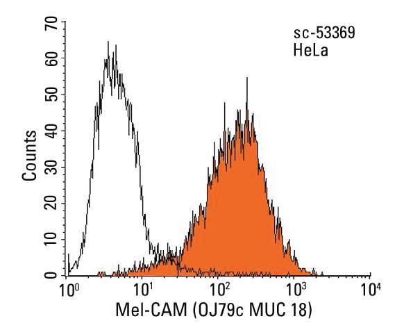
Mel-CAM (OJ79c MUC 18) - sc-53369 from Santa Cruz Biotechnology

Prices $279.00
Sizes 200 µg/ml
Host Mouse
Clonality Monoclonal
Mel-CAM (OJ79c MUC 18): sc-53369. Indirect FCM analysis of HeLa cells stained with Mel-CAM (OJ79c MUC 18), followed by PE-conjugated goat anti-mouse IgG: sc-3738. Black line histogram represents the isotype control, normal mouse IgG<sub>1</sub>: sc-3877.

Mel-CAM (2Q401) - sc-71565 from Santa Cruz Biotechnology

Prices $279.00
Sizes 200 µg/ml
Host Mouse
Clonality Monoclonal
Mel-CAM (2Q401): sc-71565. Western blot analysis of Mel-CAM expression in HeLa whole cell lysate.

Mel-CAM (3H1869) - sc-71564 from Santa Cruz Biotechnology

Prices $279.00
Sizes 200 µg/ml
Host Mouse
Clonality Monoclonal
Mel-CAM (3H1869): sc-71564. Western blot analysis of Mel-CAM expression in SK-MEL-24 whole cell lysate.

Mel-CAM (1) - sc-135987 from Santa Cruz Biotechnology

Prices $279.00
Sizes 50 µg/0.5 ml
Host Mouse
Clonality Monoclonal
Mel-CAM (1): sc-135987. Immunofluorescence staining of human endothelial cells showing cytoplasmic localization.

Anti-CD146 antibody - ab203118 from Abcam

Prices $359.00
Sizes 100 µl
Host Rabbit
Clonality Polyclonal
Immunohistochemical analysis of formalin/PFA-fixed paraffin-embedded rat brain tissue sections labeling CD146 with ab203118 at a 1/400 dilution, followed by conjugation to the secondary antibody and DAB staining.

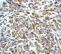
Anti-CD146 antibody - ab174326 from Abcam

Prices $384.00
Sizes 400 µl
Host Rabbit
Clonality Polyclonal
Anti-CD146 antibody (ab174326) at 1/100 dilution + A2058 cell line lysate at 35 µg

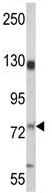
Anti-CD146 antibody - ab87342 from Abcam

Prices $376.00
Sizes 100 µg
Host Rabbit
Clonality Polyclonal
Anti-CD146 antibody (ab87342) at 1 µg/ml + Liver (Human) Tissue Lysate - adult normal tissue (ab29889) at 10 µgSecondaryGoat Anti-Rabbit IgG H&L (HRP) preadsorbed (ab97080) at 1/5000 dilutiondeveloped using the ECL techniquePerformed under reducing conditions.

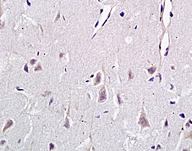
Anti-CD146 antibody - ab28360 from Abcam

Prices $378.00
Sizes 100 µg
Host Rabbit
Clonality Polyclonal

Anti-CD146 antibody [6C3F1] - ab181769 from Abcam

Prices $384.00
Sizes 100 µg
Host Mouse
Clonality Monoclonal
Clone 6C3F1
Anti-CD146 antibody [6C3F1] (ab181769) at 1/500 dilution + HeLa cell lysate

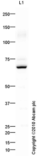
Anti-CD146 antibody [EPR3207(2)] - ab134065 from Abcam

Prices $381.00
Sizes 100 µl
Host Rabbit
Clonality Monoclonal
Clone EPR3207(2)
All lanes : Anti-CD146 antibody [EPR3207(2)] (ab134065) at 1/1000 dilutionLane 1 : A375 cell lysateLane 2 : Fetal artery lysateLane 3 : HUVEC cell lysateLysates/proteins at 10 µg per lane.SecondaryHRP labelled goat anti-rabbit at 1/2000 dilution

Anti-CD146 antibody [EPR3208] - ab75769 from Abcam

Prices $401.00
Sizes 100 µl
Host Rabbit
Clonality Monoclonal
Clone EPR3208
ab75769 at 1/250 dilution staining CD146 in melanoma tissue, using a HRP/AP polymerized secondary antibody.

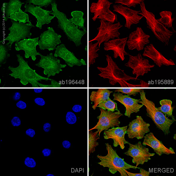
Anti-CD146 antibody [EPR3208] (Alexa Fluor® 488) - ab196448 from Abcam

Prices $425.00
Sizes 100 µl
Host Rabbit
Clonality Monoclonal
Clone EPR3208
ab196448 staining CD146 in A375 cells. The cells were fixed with 100% methanol (5 min), permeabilized with 0.1% Triton X-100 for 5 minutes and then blocked with 1% BSA/10% normal goat serum/0.3M glycine in 0.1% PBS-Tween for 1h. The cells were then incubated overnight at +4°C with ab196448 at 1/100 dilution (shown in green) and ab195889, Mouse monoclonal to alpha Tubulin (Alexa Fluor® 594), at 1/250 dilution (shown in red). Nuclear DNA was labelled with DAPI (shown in blue).Image was taken with a confocal microscope (Leica-Microsystems, TCS SP8).

Anti-CD146 antibody [N1238] - ab49492 from Abcam

Prices $378.00
Sizes 500 µl
Host Mouse
Clonality Monoclonal
Clone N1238
Ab49492 at 1/25 dilution staining metastatic melanocytes and endothelial cells from Human malignant melanoma; paraffin embedded; high temperature antigen unmasking technique was used.

Anti-CD146 antibody [OJ79c] - ab22769 from Abcam

Prices $372.00
Sizes 100 µg
Host Mouse
Clonality Monoclonal
Clone OJ79c

Anti-CD146 antibody [OJ79c] (FITC) - ab33300 from Abcam

Prices $372.00
Sizes 50 µg
Host Mouse
Clonality Monoclonal
Clone OJ79c

Anti-CD146 antibody [OJ79c MUC 18] - ab10816 from Abcam

Prices $359.00
Sizes 100 µg
Host Mouse
Clonality Monoclonal
Clone OJ79c MUC 18

Anti-CD146 antibody [P1H12] - ab24577 from Abcam

Prices $403.00
Sizes 100 µg
Host Mouse
Clonality Monoclonal
Clone P1H12
IHC image of CD146 staining in human aorta formalin fixed paraffin embedded tissue section, performed on a Leica BondTM system using the standard protocol F. The section was pre-treated using heat mediated antigen retrieval with sodium citrate buffer (pH6, epitope retrieval solution 1) for 20 mins. The section was then incubated with ab24577, 5µg/ml, for 15 mins at room temperature and detected using an HRP conjugated compact polymer system. DAB was used as the chromogen. The section was then counterstained with haematoxylin and mounted with DPX. For other IHC staining systems (automated and non-automated) customers should optimize variable parameters such as antigen retrieval conditions, primary antibody concentration and antibody incubation times.
Search more