Anti-CD45 antibody
| Name | Anti-CD45 antibody |
|---|---|
| Supplier | Abcam |
| Catalog | ab10558 |
| Prices | $400.00 |
| Sizes | 100 µg |
| Host | Rabbit |
| Clonality | Polyclonal |
| Isotype | IgG |
| Applications | FC IHC-F WB IHC-P ICC/IF ICC/IF |
| Species Reactivities | Mouse, Rat, Human, Pig, Monkey |
| Antigen | Synthetic peptide corresponding to Human CD45 aa 900-1000 conjugated to Keyhole Limpet Haemocyanin (KLH) |
| Blocking Peptide | Human CD45 peptide |
| Description | Rabbit Polyclonal |
| Gene | PTPRC |
| Conjugate | Unconjugated |
| Supplier Page | Shop |
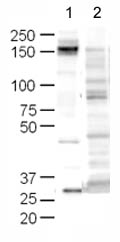
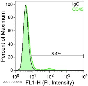
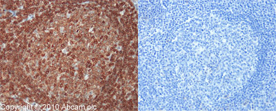
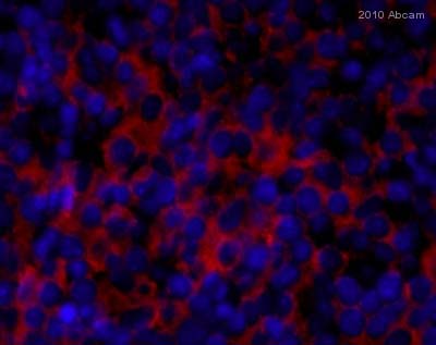
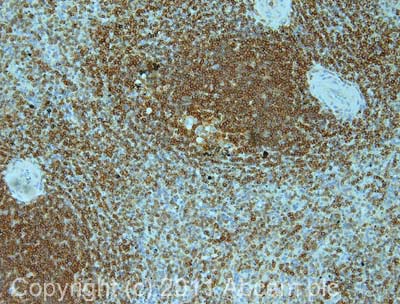
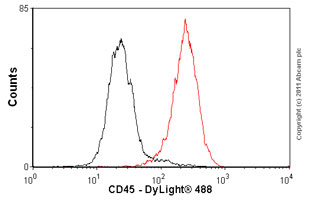
Product images
Product References
Glucose transporter expression in eutopic endometrial tissue and ectopic - Glucose transporter expression in eutopic endometrial tissue and ectopic
McKinnon B, Bertschi D, Wotzkow C, Bersinger NA, Evers J, Mueller MD. J Mol Endocrinol. 2014 Feb 24;52(2):169-79.
Bone morphogenetic protein signaling protects against cerulein-induced pancreatic - Bone morphogenetic protein signaling protects against cerulein-induced pancreatic
Gao X, Cao Y, Staloch DA, Gonzales MA, Aronson JF, Chao C, Hellmich MR, Ko TC. PLoS One. 2014 Feb 21;9(2):e89114.
Contribution of intimal smooth muscle cells to cholesterol accumulation and - Contribution of intimal smooth muscle cells to cholesterol accumulation and
Allahverdian S, Chehroudi AC, McManus BM, Abraham T, Francis GA. Circulation. 2014 Apr 15;129(15):1551-9.
Isolation and characterization of mesenchymal progenitor cells from human orbital - Isolation and characterization of mesenchymal progenitor cells from human orbital
Chen SY, Mahabole M, Horesh E, Wester S, Goldberg JL, Tseng SC. Invest Ophthalmol Vis Sci. 2014 Jul 3;55(8):4842-52.
Sonic hedgehog mediates the proliferation and recruitment of transformed - Sonic hedgehog mediates the proliferation and recruitment of transformed
Donnelly JM, Chawla A, Houghton J, Zavros Y. PLoS One. 2013 Sep 19;8(9):e75225.
Isolation and analysis of rare cells in the blood of cancer patients using a - Isolation and analysis of rare cells in the blood of cancer patients using a
Wu Y, Deighan CJ, Miller BL, Balasubramanian P, Lustberg MB, Zborowski M, Chalmers JJ. Methods. 2013 Dec 1;64(2):169-82.
Targeted disruption of MCPIP1/Zc3h12a results in fatal inflammatory disease. - Targeted disruption of MCPIP1/Zc3h12a results in fatal inflammatory disease.
Miao R, Huang S, Zhou Z, Quinn T, Van Treeck B, Nayyar T, Dim D, Jiang Z, Papasian CJ, Eugene Chen Y, Liu G, Fu M. Immunol Cell Biol. 2013 May;91(5):368-76.
Acetylsalicylic acid treatment until surgery reduces oxidative stress and - Acetylsalicylic acid treatment until surgery reduces oxidative stress and
Berg K, Langaas M, Ericsson M, Pleym H, Basu S, Nordrum IS, Vitale N, Haaverstad R. Eur J Cardiothorac Surg. 2013 Jun;43(6):1154-63.
Placental expression of CD100, CD72 and CD45 is dysregulated in human - Placental expression of CD100, CD72 and CD45 is dysregulated in human
Lorenzi T, Turi A, Lorenzi M, Paolinelli F, Mancioli F, La Sala L, Morroni M, Ciarmela P, Mantovani A, Tranquilli AL, Castellucci M, Marzioni D. PLoS One. 2012;7(5):e35232.
Inflamed tumor-associated adipose tissue is a depot for macrophages that - Inflamed tumor-associated adipose tissue is a depot for macrophages that
Wagner M, Bjerkvig R, Wiig H, Melero-Martin JM, Lin RZ, Klagsbrun M, Dudley AC. Angiogenesis. 2012 Sep;15(3):481-95.
