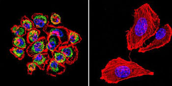
Immunofluorescent analysis of A431 cells, labeling CDC42 with ab155940 at 1/20 dilution. F-Actin staining with Phalloidin (red) and nuclei with DAPI (blue) is also shown.

Immunofluorescent analysis of C6 cells, labeling CDC42 with ab155940 at 1/20 dilution. F-Actin staining with Phalloidin (red) and nuclei with DAPI (blue) is also shown.
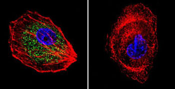
Immunofluorescent analysis of HepG2 cells, labeling CDC42 with ab155940 at 1/200 dilution. F-Actin staining with Phalloidin (red) and nuclei with DAPI (blue) is also shown.
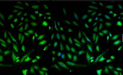
Immunofluorescent analysis of HeLa cells, labeling CDC42 with ab155940 at 1/100 dilution. Blue: Nuclei stained with Hoechst 33342.
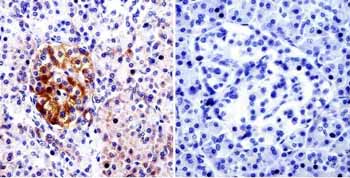
Immunohistochemical analysis of deparaffinised Human pancreas tissue, labeling CDC42 with ab155940 at 1/20 dilution. Detection was performed using a biotin conjugated secondary antibody and SA-HRP. Right image is immunohistochemistry performed without primary antibody (negative control).
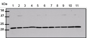
All lanes : Anti-CDC42 antibody (ab155940) at 1/1000 dilutionLane 1 : HeLa whole cell lysateLane 2 : HepG2 whole cell lysateLane 3 : 293T whole cell lysateLane 4 : A431 whole cell lysateLane 5 : Jurkat whole cell lysateLane 6 : MCF7 whole cell lysateLane 7 : U2-OS whole cell lysateLane 8 : K652 whole cell lysateLane 9 : COS7 whole cell lysateLane 10 : MEF whole cell lysateLane 11 : 3T3L1 whole cell lysateLysates/proteins at 50 µg per lane.