Anti-CEBP Beta antibody [A16]
| Name | Anti-CEBP Beta antibody [A16] |
|---|---|
| Supplier | Abcam |
| Catalog | ab18336 |
| Host | Mouse |
| Clonality | Monoclonal |
| Isotype | IgG1 |
| Clone | A16 |
| Applications | FC IHC-P IHC-F WB ICC/IF ICC/IF IHC-F |
| Species Reactivities | Mouse, Human |
| Antigen | Recombinant full length protein corresponding to Mouse CEBP Beta aa 1-268 |
| Description | Mouse Monoclonal |
| Gene | CEBPB |
| Conjugate | Unconjugated |
| Supplier Page | Shop |
Product images
Product References
Age-associated change of C/EBP family proteins causes severe liver injury and - Age-associated change of C/EBP family proteins causes severe liver injury and
Hong IH, Lewis K, Iakova P, Jin J, Sullivan E, Jawanmardi N, Timchenko L, Timchenko N. J Biol Chem. 2014 Jan 10;289(2):1106-18.
Inhibition of CD200R1 expression by C/EBP beta in reactive microglial cells. - Inhibition of CD200R1 expression by C/EBP beta in reactive microglial cells.
Dentesano G, Straccia M, Ejarque-Ortiz A, Tusell JM, Serratosa J, Saura J, Sola C. J Neuroinflammation. 2012 Jul 9;9:165.
Role of C/EBPbeta transcription factor in adult hippocampal neurogenesis. - Role of C/EBPbeta transcription factor in adult hippocampal neurogenesis.
Cortes-Canteli M, Aguilar-Morante D, Sanz-Sancristobal M, Megias D, Santos A, Perez-Castillo A. PLoS One. 2011;6(10):e24842.
CCAAT/enhancer binding protein beta deficiency provides cerebral protection - CCAAT/enhancer binding protein beta deficiency provides cerebral protection
Cortes-Canteli M, Luna-Medina R, Sanz-Sancristobal M, Alvarez-Barrientos A, Santos A, Perez-Castillo A. J Cell Sci. 2008 Apr 15;121(Pt 8):1224-34.


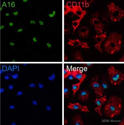
 used under the same conditions. Acquisition of >5,000 events was performed. This antibody gave a positive signal in HT1080 cells fixed with 4% paraformaldehyde/permeabilized in 0.1% PBS-Tween used under the same conditions.](http://www.bioprodhub.com/system/product_images/ab_products/2/sub_1/27372_CEBP-Beta-Primary-antibodies-ab18336-4.jpg)