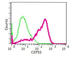
Overlay histogram showing HepG2 cells stained with ab170414 (red line) at 1/170 dilution. The cells were fixed with 2% paraformaldehyde. The secondary antibody used was a FITC conjugated goat anti-rabbit IgG at 1/150 dilution. Isotype control antibody (green line) was rabbit monoclonal IgG used under the same conditions.
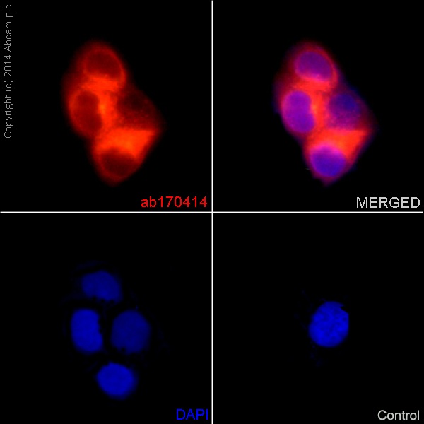
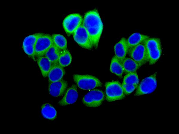
ab170414 staining CEP55 in the HepG2 cell line by ICC/IF (Immunocytochemistry/immunofluorescence). Cells were fixed with 4% Paraformaldehyde. Samples were incubated with primary antibody (1/400). An Alexa Fluor®555-conjugated Goat anti-rabbit IgG(1/500) was used as the secondary antibody. Nuclei were counterstained with DAPI.
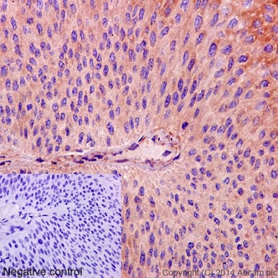
ab170414 staining CEP55 in Human transitional cell carcinoma of bladder tissue sections by Immunohistochemistry (IHC-P - paraformaldehyde-fixed, paraffin-embedded sections). Tissue was fixed and paraffin-embedded, antigen retrieval was by heat mediation in Tris/EDTA buffer pH9. Samples were incubated with primary antibody (1/400). An undiluted HRP-conjugated anti-rabbit IgG was used as the secondary antibody. Tissue counterstained with Hematoxylin. PBS was used in the negative control rather than the Primary antibody.
![Anti-CEP55 antibody [EPR11944(B)] (ab170414) at 1/5000 dilution + SW480 Cell Lysate at 10 µgSecondaryGoat Anti-Rabbit IgG, (H+L), HRP-conjugated at 1/1000 dilution](http://www.bioprodhub.com/system/product_images/ab_products/2/sub_1/27771_ab170414-240550-170414-WB2.jpg)
Anti-CEP55 antibody [EPR11944(B)] (ab170414) at 1/5000 dilution + SW480 Cell Lysate at 10 µgSecondaryGoat Anti-Rabbit IgG, (H+L), HRP-conjugated at 1/1000 dilution
![Anti-CEP55 antibody [EPR11944(B)] (ab170414) at 1/20000 dilution + HeLa cell lysate at 10 µgSecondaryGoat Anti-Rabbit IgG, (H+L), HRP-conjugated at 1/1000 dilution](http://www.bioprodhub.com/system/product_images/ab_products/2/sub_1/27772_ab170414-240547-170414-WB1.jpg)
Anti-CEP55 antibody [EPR11944(B)] (ab170414) at 1/20000 dilution + HeLa cell lysate at 10 µgSecondaryGoat Anti-Rabbit IgG, (H+L), HRP-conjugated at 1/1000 dilution
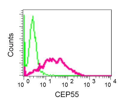
Flow cytometric analysis of permeabilized CEP55 cells labeling CEP55 with ab170414, unpurified (red) or a rabbit IgG (negative) (green).
Immunofluorescence analysis of HeLa cells, labeling CEP55 using ab170414, unpurified.
![All lanes : Anti-CEP55 antibody [EPR11944(B)] (ab170414) at 1/20000 dilution (unpurified)Lane 1 : SW480 cell lysateLane 2 : HeLa cell lysateLane 3 : HepG2 cell lysatedeveloped using the ECL techniquePerformed under reducing conditions.](http://www.bioprodhub.com/system/product_images/ab_products/2/sub_1/27775_ab170414-169414-78251wbAbcamAdmin.jpg)
All lanes : Anti-CEP55 antibody [EPR11944(B)] (ab170414) at 1/20000 dilution (unpurified)Lane 1 : SW480 cell lysateLane 2 : HeLa cell lysateLane 3 : HepG2 cell lysatedeveloped using the ECL techniquePerformed under reducing conditions.