Anti-Clathrin antibody [X22]
| Name | Anti-Clathrin antibody [X22] |
|---|---|
| Supplier | Abcam |
| Catalog | ab2731 |
| Prices | $403.00 |
| Sizes | 100 µl |
| Host | Mouse |
| Clonality | Monoclonal |
| Isotype | IgG1 |
| Clone | X22 |
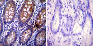
| Applications | ICC/IF ICC/IF ICC/IF ELISA B/N FC Immunomicroscopy IP WB IHC-P |
| Species Reactivities | Mouse, Rat, Bovine, Dog, Human, Pig, Xenopus, Bird, Monkey |
| Antigen | Full length native protein (purified) corresponding to Human Clathrin |
| Description | Mouse Monoclonal |
| Gene | AP1B1 |
| Conjugate | Unconjugated |
| Supplier Page | Shop |
Product images
Product References
Disruption of the coxsackievirus and adenovirus receptor-homodimeric interaction - Disruption of the coxsackievirus and adenovirus receptor-homodimeric interaction
Salinas S, Zussy C, Loustalot F, Henaff D, Menendez G, Morton PE, Parsons M, Schiavo G, Kremer EJ. J Biol Chem. 2014 Jan 10;289(2):680-95.
The adaptor protein GULP promotes Jedi-1-mediated phagocytosis through a - The adaptor protein GULP promotes Jedi-1-mediated phagocytosis through a
Sullivan CS, Scheib JL, Ma Z, Dang RP, Schafer JM, Hickman FE, Brodsky FM, Ravichandran KS, Carter BD. Mol Biol Cell. 2014 Jun 15;25(12):1925-36.
Deficits in receptor-mediated endocytosis and recycling in cells from mice with - Deficits in receptor-mediated endocytosis and recycling in cells from mice with
Zhou GL, Na SY, Niedra R, Seed B. J Cell Sci. 2014 Sep 15;127(Pt 18):3916-27.
A huntingtin-mediated fast stress response halting endosomal trafficking is - A huntingtin-mediated fast stress response halting endosomal trafficking is
Nath S, Munsie LN, Truant R. Hum Mol Genet. 2015 Jan 15;24(2):450-62.
Profilin 1 as a target for cathepsin X activity in tumor cells. - Profilin 1 as a target for cathepsin X activity in tumor cells.
Pecar Fonovic U, Jevnikar Z, Rojnik M, Doljak B, Fonovic M, Jamnik P, Kos J. PLoS One. 2013;8(1):e53918.
Glucocorticoid receptor beta and histone deacetylase 1 and 2 expression in the - Glucocorticoid receptor beta and histone deacetylase 1 and 2 expression in the
Butler CA, McQuaid S, Taggart CC, Weldon S, Carter R, Skibinski G, Warke TJ, Choy DF, McGarvey LP, Bradding P, Arron JR, Heaney LG. Thorax. 2012 May;67(5):392-8.
Endocytosis regulates cell soma translocation and the distribution of adhesion - Endocytosis regulates cell soma translocation and the distribution of adhesion
Shieh JC, Schaar BT, Srinivasan K, Brodsky FM, McConnell SK. PLoS One. 2011 Mar 22;6(3):e17802.
Effect of electromagnetic field on endocytosis of cationic solid lipid - Effect of electromagnetic field on endocytosis of cationic solid lipid
Kuo YC, Chen HH. J Drug Target. 2010 Jul;18(6):447-56.
Quantitative proteomics identifies a Dab2/integrin module regulating cell - Quantitative proteomics identifies a Dab2/integrin module regulating cell
Teckchandani A, Toida N, Goodchild J, Henderson C, Watts J, Wollscheid B, Cooper JA. J Cell Biol. 2009 Jul 13;186(1):99-111.
Myosin VI undergoes cargo-mediated dimerization. - Myosin VI undergoes cargo-mediated dimerization.
Yu C, Feng W, Wei Z, Miyanoiri Y, Wen W, Zhao Y, Zhang M. Cell. 2009 Aug 7;138(3):537-48.
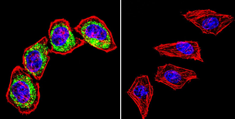
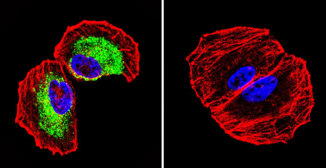
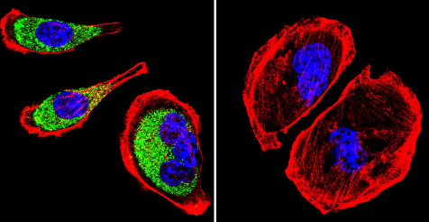
![All lanes : Anti-Clathrin antibody [X22] (ab2731) at 1/500 dilutionLane 1 : HepG2 (Human hepatocellular liver carcinoma cell line) Whole Cell Lysate Lane 2 : Raji (Human Burkitt's lymphoma cell line) Whole Cell Lysate Lysates/proteins at 10 µg per lane.SecondaryGoat polyclonal to Mouse IgG - H&L - Pre-Adsorbed (HRP) at 1/3000 dilutionObserved band size : 180 kDa (why is the actual band size different from the predicted?)Additional bands at : 240 kDa,450 kDa. We are unsure as to the identity of these extra bands.](http://www.bioprodhub.com/system/product_images/ab_products/2/sub_1/29263_Clathrin-Primary-antibodies-ab2731-2.jpg)
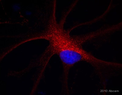
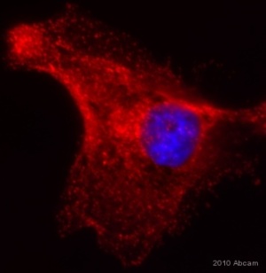
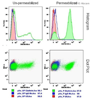
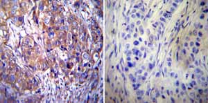
![Xenopus laevis cytoplasmic egg extract visualized live with primary and secondary antibody addition [red is anti-Clathrin X22 (ab2731) with goat anti-mouse Alexa Fluor 568 secondary, green is anti-HIP1R (ab77297) with goat anti-rabbit Alexa Fluor 488 secondary]. Large red structures are probably aggregates, but the small structures appear to be specific for vesicle staining.See Abreview](http://www.bioprodhub.com/system/product_images/ab_products/2/sub_1/29269_Clathrin-Primary-antibodies-ab2731-59.jpg)