Anti-CLLD8/SETDB2 antibody (ab5517) at 1 µg/ml + Human CLLD8/SETDB2 full length protein (ab169554) at 0.01 µgSecondaryGoat Anti-Rabbit IgG H&L (HRP) (ab97051) at 1/10000 dilutiondeveloped using the ECL techniquePerformed under reducing conditions.Additional bands at : 120 kDa (possible tagged protein).Exposure time : 10 secondsab5517 recognises a band corresponding to the tagged CLLD8/SETB2 at approximately 120 kDa. Abcam recommends using milk as the blocking agent. Abcam welcomes customer feedback and would appreciate any comments regarding this product and the data presented above.

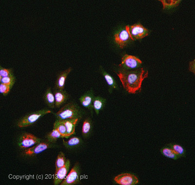
ICC/IF image of ab5517 stained MCF-7 cells. The cells were 4% formaldehyde fixed (10 min) and then incubated in 1%BSA / 10% normal Goat serum / 0.3M glycine in 0.1% PBS-Tween for 1h to permeabilise the cells and block non-specific protein-protein interactions. The cells were then incubated with the antibody ab5517 at 5ug/ml overnight at +4°C. The secondary antibody (green) was DyLight® 488 Goat anti Rabbit (ab96899) IgG (H+L) used at a 1/250 dilution for 1h. Alexa Fluor® 594 WGA was used to label plasma membranes (red) at a 1/200 dilution for 1h. DAPI was used to stain the cell nuclei (blue) at a concentration of 1.43µM.

Left: Endogenous CLLD8Detection using indirect fluorescence of the signal corresponding to endogenous CLLD8 in 293T cells. Cells fixed using 4% formaldehyde, blocked with PBS containing 3% milk and 0.5% Triton X-100, incubated for 1 hour at 37 °C using a 1/50 dilution of antibody ab5517. Cells were then washed 3 times and incubated for 1 hour at 37 °C with a goat anti-rabbit secondary antibody (1/500 dilution) coupled to Alexa Fluor 555 (Molecular Probes). Following 3 more washes, cells were stained with DAPI and mounted with Vectashield.Top: DAPIBottom: ab5517Right: Overexpressed CLLD8Detection using indirect fluorescence of the signal corresponding to staining with anti-FLAG mouse antibody (top) and an antibody (ab5517) against CLLD8 (middle) in 293T cells. 293T cells were transfected with vectors for overexpression of flagged CLLD8 using Lipofectamine 2000 transfection procedure (Invitrogen). Cells were fixed 48 hours post-tra

Detection using indirect fluorescence of the signal corresponding to endogenous CLLD8 in 293T cells. Using Lipofectamine 2000 (Invitrogen), cells were transfected with either a control shRNA directed against luciferase (left, SiRluc) or a specific siRNA directed against CLLD8 (right). 48 hours post-transfection, cells were fixed, blocked, and incubated with a 1:50 dilution of ab5517 at 37 °C for 1 hour. Cells were then washed 3 times and incubated at 37 °C for 1 hour with a 1/500 dilution of goat anti-rabbit antibody coupled with Alexa Fluor 555 (Molecular Probes). Following 3 further washes cells were stained with DAPI and mounted with Vectashield. Antibody signals were extinguished when a specific RNAi was used. The few cells which retained a signal were presumably not transfected.

All lanes : Anti-CLLD8/SETDB2 antibody (ab5517) at 1 µg/mlLane 1 : Sf21 cells infected by baculovirus containing FLAG-CLLD8 gene at 10 µgLane 2 : Sf21 cells infected by baculovirus containing FLAG-CLLD8 gene at 20 µgLane 3 : non-infected Sf21 cells at 10 µgLane 4 : non-infected Sf21 cells at 20 µgLane 5 : Sf21 infected by baculovirus containing FLAG-CLLD8 gene at 10 µg with Human CLLD8/SETDB2 peptide (ab24398) at 1 µg/mlLane 6 : Sf21 infected by baculovirus containing FLAG-CLLD8 gene at 20 µg with Human CLLD8/SETDB2 peptide (ab24398) at 1 µg/mlLane 7 : non-infected Sf21 cells at 10 µg with Human CLLD8/SETDB2 peptide (ab24398) at 1 µg/mlLane 8 : non-infected Sf21 cells at 20 µg with Human CLLD8/SETDB2 peptide (ab24398) at 1 µg/mlObserved band size : 110 kDa (why is the actual band size different from the predicted?)Additional bands at : 22 kDa (possible non-specific binding),90 kDa (possible non-specific binding).ab5517 recognises a band corresponding to FLAG-tagged CLLD8/SETB2 at appro

Image courtesy of Human Protein Atlas ab5517 staining CLLD8 in human colon tissue and showing a distinct and strong staining of the nucleus of the glandular cells. Paraffin embedded human colon was incubated with ab5517 (1/50) for 30 minutes at room temperature. Antigen retrieval was performed by heat induction in citrate buffer pH6. ab5517 was tested in a tissue microarray (TMA) containing a wide range of normal and cancer tissues as well as a cell microarray consisting of a range of commonly used, well characterised human cell lines. Further results for this antibody can be found at www.proteinatlas.org


