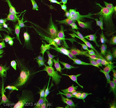
ICC/IF image of ab79261 stained HepG2 cells. The cells were 4% formaldehyde fixed (10 min) and then incubated in 1%BSA / 10% normal goat serum / 0.3M glycine in 0.1% PBS-Tween for 1h to permeabilise the cells and block non-specific protein-protein interactions. The cells were then incubated with the antibody ab79261 at 1/1000 dilution overnight at +4°C. The secondary antibody (green) was DyLight® 488 Goat anti-Rabbit IgG (H+L) (ab96899) used at a 1/250 dilution for 1h. Alexa Fluor® 594 WGA was used to label plasma membranes (red) at a 1/200 dilution for 1h. DAPI was used to stain the cell nuclei (blue) at a concentration of 1.43µM.
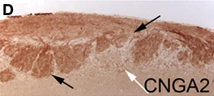
ab79261, staining CNGA2 in murine olfactory bulb tissue by Immunohistochemistry. Olfactory bulbs from CNGA2 heterozygous mice were fixed in 10% neutral buffered formalin overnight and embedded in paraffin. Sections were deparaffinized and microwaved in antigen retrieval buffer (10 mM Tris pH 9.0, 1 mM EDTA, 0.05% Tween 20). Slides were blocked in TNB before incubation with primary antibody overnight at 4°C. A biotinylated goat anti-rabbit IgG was used as the secondary antibody.