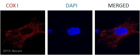
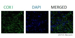
Anti-COX1 / Cyclooxygenase 1 antibody [5F6/F4]
| Name | Anti-COX1 / Cyclooxygenase 1 antibody [5F6/F4] |
|---|---|
| Supplier | Abcam |
| Catalog | ab695 |
| Prices | $403.00 |
| Sizes | 50 µg |
| Host | Mouse |
| Clonality | Monoclonal |
| Isotype | IgG1 |
| Clone | 5F6/F4 |
| Applications | ELISA WB IHC-F IHC-P FC IP ICC/IF ICC/IF |
| Species Reactivities | Mouse, Rat, Human |
| Antigen | A preparation of intracellular proteins of the human cell line HL60 |
| Description | Mouse Monoclonal |
| Gene | PTGS1 |
| Conjugate | Unconjugated |
| Supplier Page | Shop |
Product images
Product References
Low-dose aspirin delays an inflammatory tumor progression in vivo in a transgenic - Low-dose aspirin delays an inflammatory tumor progression in vivo in a transgenic
Carlson LM, Rasmuson A, Idborg H, Segerstrom L, Jakobsson PJ, Sveinbjornsson B, Kogner P. Carcinogenesis. 2013 May;34(5):1081-8.
Flaxseed enriched diet-mediated reduction in ovarian cancer severity is - Flaxseed enriched diet-mediated reduction in ovarian cancer severity is
Eilati E, Hales K, Zhuge Y, Ansenberger Fricano K, Yu R, van Breemen RB, Hales DB. Prostaglandins Leukot Essent Fatty Acids. 2013 Sep;89(4):179-87. doi:
Serotonin 2B receptor (5-HT2B R) signals through prostacyclin and PPAR-ss/delta - Serotonin 2B receptor (5-HT2B R) signals through prostacyclin and PPAR-ss/delta
Chabbi-Achengli Y, Launay JM, Maroteaux L, de Vernejoul MC, Collet C. PLoS One. 2013 Sep 17;8(9):e75783.
Interaction of cyclooxygenases with an apoptosis- and autoimmunity-associated - Interaction of cyclooxygenases with an apoptosis- and autoimmunity-associated
Ballif BA, Mincek NV, Barratt JT, Wilson ML, Simmons DL. Proc Natl Acad Sci U S A. 1996 May 28;93(11):5544-9.


![Anti-COX1 / Cyclooxygenase 1 antibody [5F6/F4] (ab695) at 1 µg/ml + whole cell lysate prepared from human huh-7 cell line at 15 µgSecondarySheep anti-mouse IgG conjugated to HRP at 1/1000 dilutionPerformed under reducing conditions.Observed band size : 68 kDa (why is the actual band size different from the predicted?)Additional bands at : 38 kDa. We are unsure as to the identity of these extra bands.Exposure time : 4 minutesThis image is courtesy of an anonymous abreview.Primary antibody incubated for 1 hour at 20°C in 5% milk in TBST.Gel running conditions: Denaturing.Blocked using 5% milk for 16 hours at 4°C.Detection method: Western lightning chemiluminescent reagent.See Abreview](http://www.bioprodhub.com/system/product_images/ab_products/2/sub_2/1308_COX1-Cyclooxygenase-1-Primary-antibodies-ab695-3.jpg)
![Overlay histogram showing NIH3T3 cells stained with ab695 (red line). The cells were fixed with 80% methanol (5 min) and then permeabilized with 0.1% PBS-Tween for 20 min. The cells were then incubated in 1x PBS / 10% normal goat serum / 0.3M glycine to block non-specific protein-protein interactions followed by the antibody (ab695, 2µg/1x106 cells) for 30 min at 22ºC. The secondary antibody used was DyLight® 488 goat anti-mouse IgG (H+L) (ab96879) at 1/500 dilution for 30 min at 22ºC. Isotype control antibody (black line) was mouse IgG1 [ICIGG1] (ab91353, 2µg/1x106 cells) used under the same conditions. Acquisition of >5,000 events was performed.](http://www.bioprodhub.com/system/product_images/ab_products/2/sub_2/1309_COX1-Cyclooxygenase-1-Primary-antibodies-ab695-8.jpg)

