Anti-CRB3 antibody [14F9]
| Name | Anti-CRB3 antibody [14F9] |
|---|---|
| Supplier | Abcam |
| Catalog | ab180835 |
| Prices | $376.00 |
| Sizes | 100 µg |
| Host | Rat |
| Clonality | Monoclonal |
| Isotype | IgG2c |
| Clone | 14F9 |
| Applications | WB ICC/IF ICC/IF ICC/IF |
| Species Reactivities | Mouse, Human |
| Antigen | Full length protein corresponding to Rat CRB3 |
| Description | Rat Monoclonal |
| Gene | CRB3 |
| Conjugate | Unconjugated |
| Supplier Page | Shop |
Product images
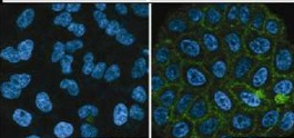 Immunofluorescence analysis of HeLa cell labeling CRB3 with ab1808350 at a 1/100 dilution. DAPI was used to stain the nuclei.
![All lanes : Anti-CRB3 antibody [14F9] (ab180835) at 1/1000 dilutionLane 1 : HEK293T cells overexpressed with control vectorLane 2 : HEK293T cells overexpressed with CRB3Lane 3 : Mouse kidney tissue lysateLane 4 : Mouse colon tissue lysate](http://www.bioprodhub.com/system/product_images/ab_products/2/sub_2/1973_ab180835-209559-ab1808352.png) All lanes : Anti-CRB3 antibody [14F9] (ab180835) at 1/1000 dilutionLane 1 : HEK293T cells overexpressed with control vectorLane 2 : HEK293T cells overexpressed with CRB3Lane 3 : Mouse kidney tissue lysateLane 4 : Mouse colon tissue lysate
All lanes : Anti-CRB3 antibody [14F9] (ab180835) at 1/1000 dilutionLane 1 : HEK293T cells overexpressed with control vectorLane 2 : HEK293T cells overexpressed with CRB3Lane 3 : Mouse kidney tissue lysateLane 4 : Mouse colon tissue lysate
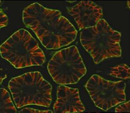 Immunofluorescence analysis of Mouse intestine labeling CRB3 with ab1808350 at a 1/100 dilution. E-cadherin staining was used to highlight the lateral membrane in mouse intestine.