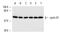
cyclin B1 (H-433): sc-752. Western blot analysis of cyclin B1 expression in untreated (A,C,E) and phorbol ester-induced (B,D,F) K-562 (A,B), Jurkat (C,D) and HeLa (E,F) nuclear extracts.
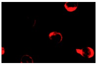
cyclin B1 (H-433): sc-752. Immunofluorescence staining of methanol-fixed K-562 cells showing cytoplasmic staining.
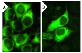
cyclin B1 siRNA (h): sc-29284. Immunofluorescence staining of methanol-fixed, control HeLa (A) and cyclin B1 siRNA silenced HeLa (B) cells showing diminished cytoplasmic staining in the siRNA silenced cells. Cells probed with cyclin B1 (H-433): sc-752.
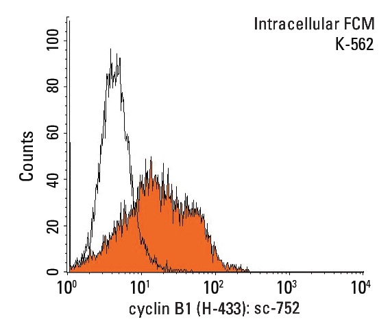
cyclin B1 (H-433): sc-752. Indirect, intracellular FCM analysis of fixed and permeabilized K-562 cells stained with cyclin B1 (H-433), followed by PE-conjugated goat anti-rabbit IgG: sc-3739. Black line histogram represents the isotype control, normal rabbit IgG: sc-3888.
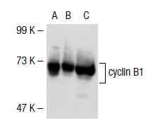
cyclin B1 (H-433): sc-752. Western blot analysis of cyclin B1 expression in non-transfected: sc-117752 (A) and mouse cyclin B1 transfected: sc-119543 (B) 293T whole cell lysates and K-562 nuclear extract (C).
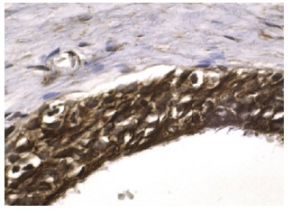
cyclin B1 (H-433): sc-752. Immunoperoxidase staining of formalin fixed, paraffin-embedded human testis tissue showing nuclear and cytoplasmic staining of cells in seminiferus ducts.
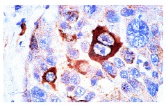
cyclin B1 (H-433): sc-752. Immunoperoxidase staining of formalin-fixed, paraffin-embedded human breast carcinoma tissue at high magnification. Note staining of selected cells, showing cytoplasmic localization.
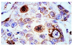
cyclin B1 (H-433): sc-752. Immunoperoxidase staining of formalin-fixed, paraffin-embedded human breast carcinoma tissue at high magnification. Note staining of selected cells, showing nuclear localization.
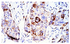
cyclin B1 (H-433): sc-752. Immunoperoxidase staining of formalin-fixed, paraffin-embedded human breast carcinoma tissue at low magnification.


