Anti-Cytochrome P450 4A antibody
| Name | Anti-Cytochrome P450 4A antibody |
|---|---|
| Supplier | Abcam |
| Catalog | ab3573 |
| Prices | $390.00 |
| Sizes | 100 µl |
| Host | Rabbit |
| Clonality | Polyclonal |
| Isotype | IgG |
| Applications | IHC-F WB ICC/IF ICC/IF IHC-P IP |
| Species Reactivities | Mouse, Rat, Rabbit, Hamster, Cat, Dog, Human, Pig |
| Antigen | Synthetic peptide corresponding to Rat Cytochrome P450 4A aa 431-445 |
| Description | Rabbit Polyclonal |
| Gene | CYP4A11 |
| Conjugate | Unconjugated |
| Supplier Page | Shop |
Product images
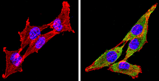 ab3573 labelling Cytochrome P450 4A (green) in the cytoplasm and membrane of H-4-II-E cells (right), compared to control (left), by Immunocytochemistry/Immunofluorescence. Formalin-fixed cells were permeabilized with 0.1% Triton X-100 in TBS for 5-10 minutes and blocked with 3% BSA-PBS for 30 minutes at room temperature. Cells were incubated with the primary antibody (1:100 in 3% BSA-PBS) overnight at 4 ºC. A DyLight-conjugated anti-rabbit was used as the secondary antibody. Red (phalloidin) - F-actin, Blue - nuclei. Images were taken at a magnification of 60x.
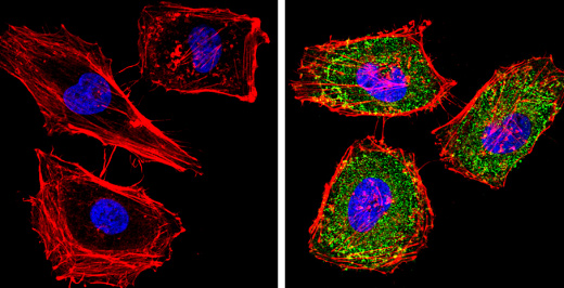 ab3573 labelling Cytochrome P450 4A (green) in the cytoplasm and membrane of HeLa cells (right), compared to control (left), by Immunocytochemistry/Immunofluorescence. Formalin-fixed cells were permeabilized with 0.1% Triton X-100 in TBS for 5-10 minutes and blocked with 3% BSA-PBS for 30 minutes at room temperature. Cells were incubated with the primary antibody (1:100 in 3% BSA-PBS) overnight at 4 ºC. A DyLight-conjugated anti-rabbit was used as the secondary antibody. Red (phalloidin) - F-actin, Blue - nuclei. Images were taken at a magnification of 60x.
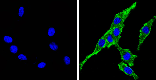 ab3573 labelling Cytochrome P450 4A (green) in the cytoplasm and membrane of PC12 cells (right), compared to control (left), by Immunocytochemistry/Immunofluorescence. Formalin-fixed cells were permeabilized with 0.1% Triton X-100 in TBS for 5-10 minutes and blocked with 3% BSA-PBS for 30 minutes at room temperature. Cells were incubated with the primary antibody (1:100 in 3% BSA-PBS) overnight at 4 ºC. A DyLight-conjugated anti-rabbit was used as the secondary antibody. Red (phalloidin) - F-actin, Blue - nuclei. Images were taken at a magnification of 60x.
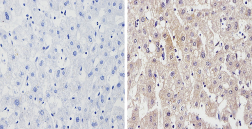
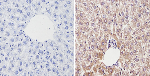 ab3573 labelling Cytochrome P450 4A in the cytoplasm of Rat liver tissue (right) compared with a negative control in the absence of primary antibody (left). To expose target proteins, antigen retrieval method was performed using 10mM sodium citrate (pH 6.0) microwaved for 8-15 min. Tissues were blocked in 3% H2O2-methanol for 15 min at room temperature. Tissue sections were incubated with the primary antibody (1:200 in 3% BSA-PBS) overnight at 4°C. A HRP-conjugated anti-rabbit was used as the secondary antibody, followed by colorimetric detection using a DAB kit. Tissues were counterstained with hematoxylin and dehydrated with ethanol and xylene to prep for mounting.
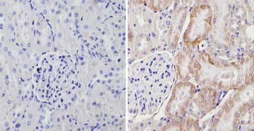 ab3573 labelling Cytochrome P450 4A in the cytoplasm of Rat kidney tissue (right) compared with a negative control in the absence of primary antibody (left). To expose target proteins, antigen retrieval method was performed using 10mM sodium citrate (pH 6.0) microwaved for 8-15 min. Tissues were blocked in 3% H2O2-methanol for 15 min at room temperature. Tissue sections were incubated with the primary antibody (1:200 in 3% BSA-PBS) overnight at 4°C. A HRP-conjugated anti-rabbit was used as the secondary antibody, followed by colorimetric detection using a DAB kit. Tissues were counterstained with hematoxylin and dehydrated with ethanol and xylene to prep for mounting.
 ab3573 labelling Cytochrome P450 4A in the cytoplasm and membrane of Human liver tissue (right) compared with a negative control in the absence of primary antibody (left). To expose target proteins, antigen retrieval method was performed using 10mM sodium citrate (pH 6.0) microwaved for 8-15 min. Tissues were blocked in 3% H2O2-methanol for 15 min at room temperature. Tissue sections were incubated with the primary antibody (1:200 in 3% BSA-PBS) overnight at 4°C. A HRP-conjugated anti-rabbit was used as the secondary antibody, followed by colorimetric detection using a DAB kit. Tissues were counterstained with hematoxylin and dehydrated with ethanol and xylene to prep for mounting.
Product References
Combined therapy with COX-2 inhibitor and 20-HETE inhibitor reduces colon tumor - Combined therapy with COX-2 inhibitor and 20-HETE inhibitor reduces colon tumor
Zhang Y, Hoda MN, Zheng X, Li W, Luo P, Maddipati KR, Seki T, Ergul A, Wang MH. Am J Physiol Regul Integr Comp Physiol. 2014 Sep 15;307(6):R693-703. doi:
Daily exercise vs. caloric restriction for prevention of nonalcoholic fatty liver - Daily exercise vs. caloric restriction for prevention of nonalcoholic fatty liver
Rector RS, Uptergrove GM, Morris EM, Borengasser SJ, Laughlin MH, Booth FW, Thyfault JP, Ibdah JA. Am J Physiol Gastrointest Liver Physiol. 2011 May;300(5):G874-83. doi:
PPAR-alpha agonist fenofibrate induces renal CYP enzymes and reduces blood - PPAR-alpha agonist fenofibrate induces renal CYP enzymes and reduces blood
Zhao X, Li LY. Am J Nephrol. 2008;28(4):598-606.
Sex hormone influence on hepatitis in young male A/JCr mice infected with - Sex hormone influence on hepatitis in young male A/JCr mice infected with
Theve EJ, Feng Y, Taghizadeh K, Cormier KS, Bell DR, Fox JG, Rogers AB. Infect Immun. 2008 Sep;76(9):4071-8.
Protective effect of 20-hydroxyeicosatetraenoic acid (20-HETE) on glomerular - Protective effect of 20-hydroxyeicosatetraenoic acid (20-HETE) on glomerular
McCarthy ET, Sharma R, Sharma M. Kidney Int. 2005 Jan;67(1):152-6.