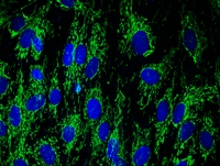
Immunocytochemistry image of ab110306-stained Human HDFn cells. The cells were paraformaldehyde fixed (4%, 20 min) and Triton X-100 permeabilized (0.1%, 15 min). Cells were then incubated with ab110306 at 1 µg/ml for 2 h at room temperature or over night at 4°C. The secondary antibody was (green) Alexa Fluor® 488 goat anti-mouse IgG (H+L) used at a 1/1000 dilution for 1 h. 10% Goat serum was used as the blocking agent for all blocking steps. DAPI was used to stain the cell nuclei (blue). Target protein locates mainly in mitochondria.

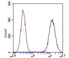
HL-60 cells were stained with 1 µg/mL ab110306 (blue) or an equal amount of an isotype control antibody (red) and analyzed by Flow Cytometry.

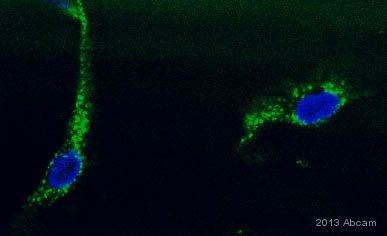
ab110306 staining DLST in Human monocyte-derived macrophages by ICC/IF (Immunocytochemistry/immunofluorescence). Cells were fixed with paraformaldehyde, permeabilized with Triton X-100 0.1% and blocked with 5% Goat serum for 60 minutes at 21°C. Samples were incubated with primary antibody (1/1000 in PBS + 1% BSA) for 12 hours at 4°C. A Cy2®-conjugated Goat anti-mouse IgG polyclonal was used as the secondary antibody.See Abreview

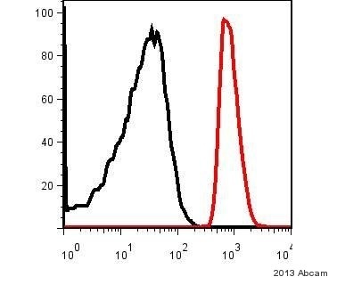
ab110306 staining DLST in Human monocyte-derived macrophages by Flow Cytometry. Cells were prepared by scraoing in PBS and fixation by paraformaldehyde. The sample was incubated with the primary antibody (1/1000 in PBS + 1% BSA) for 60 minutes at 4°C. An Alexa Fluor®488-conjugated Goat anti-mouse IgG polyclonal(1/100) was used as the secondary antibody. Gating Strategy: Dead cells excluded.See Abreview
![All lanes : Anti-DLST antibody [9F4BD5] (ab110306) at 1/4000 dilutionLane 1 : HeLa Cell lysateLane 2 : Hek293T Cell lysateLysates/proteins at 50 µg per lane.SecondaryDyLight®650-conjugated Goat anti-mouse at 1/10000 dilutiondeveloped using the ECL techniquePerformed under reducing conditions.](http://www.bioprodhub.com/system/product_images/ab_products/2/sub_2/8733_DLST-Primary-antibodies-ab110306-5.jpg)
All lanes : Anti-DLST antibody [9F4BD5] (ab110306) at 1/4000 dilutionLane 1 : HeLa Cell lysateLane 2 : Hek293T Cell lysateLysates/proteins at 50 µg per lane.SecondaryDyLight®650-conjugated Goat anti-mouse at 1/10000 dilutiondeveloped using the ECL techniquePerformed under reducing conditions.


![All lanes : Anti-DLST antibody [9F4BD5] (ab110306) at 1/4000 dilutionLane 1 : HeLa Cell lysateLane 2 : Hek293T Cell lysateLysates/proteins at 50 µg per lane.SecondaryDyLight®650-conjugated Goat anti-mouse at 1/10000 dilutiondeveloped using the ECL techniquePerformed under reducing conditions.](http://www.bioprodhub.com/system/product_images/ab_products/2/sub_2/8733_DLST-Primary-antibodies-ab110306-5.jpg)